T Cell Nomenclature Gets Its Long-Overdue Standardization! (Plus: scRNA-seq T Cell Annotation Tool Recommendations)
T cells have been back in the spotlight lately. First, regulatory T cells (Tregs) claimed this year's Nobel Prize. Then, just days ago, a landmark paper appeared in Nature proposing a unified T cell nomenclature. Today, let's break down how T cells should really be classified — and, in the single-cell world, how to annotate them properly.
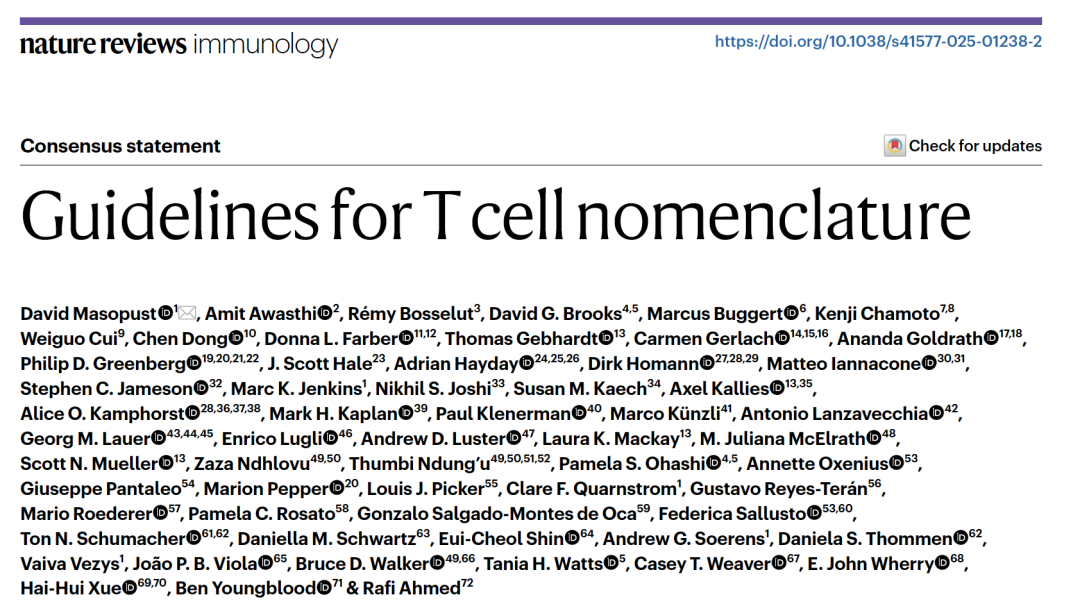 What a lineup. Source in figure
Background
In the 1990s, four-color flow cytometry was sufficient to divide T cells into two broad categories: TCM and TEM. But in the single-cell era, researchers have found that T cells form a continuous spectrum rather than a set of discrete "cell types." The result? A proliferation of inconsistent annotations: some papers name subsets by surface markers, while others coin functional names based on gene-set enrichment scores.
Where does the problem lie?
- Same term, different definitions: T
CMis defined by CCR7 in humans but by CD62L in mice; some researchers emphasize migratory capacity, others emphasize proliferative potential. - Runaway naming of new subsets: TSCM, MPEC, SLEC, LLEC, TPEX… the list keeps growing.
- Conflation of function, migration, and differentiation state: The name "TEM" bundles "effector" and "memory" into one label — so which property does it actually emphasize?
To address these issues, attendees at the 2020 Keystone Symposium on T Cell Memory put forward three recommendations.
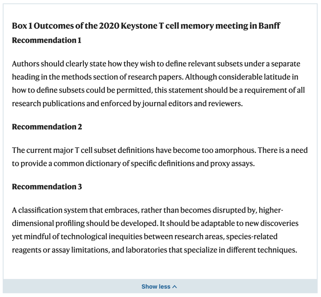 Box 1, Masopust et al., 2025
In brief:
- Methods sections must clearly state your definitions.
- There should be a standardized nomenclature "dictionary" with a corresponding marker reference table.
- There should be a classification system capable of accommodating new technologies and discoveries.
Writing Out Your Definitions
The first recommendation is directed at journals and the broader research community. Here are a few examples of how a well-structured paper should define its cell populations:
Example 1: Flow cytometry-based phenotypic definition
- Results: We compared naive CD4⁺ T cells (CD4⁺ T
N), activated CD4⁺ T cells (CD4⁺ TA), and memory CD4⁺ T cells (CD4⁺ TM).- Methods: CD4⁺ T cells were defined by flow cytometric phenotype. CD4⁺ T
Nwere defined as CD44^low^ CD62L⁺; CD4⁺ TAwere defined as CD44^hi^ CD62L⁻ with documented antigen stimulation within one week prior to analysis; CD4⁺ TMwere defined as CD44^hi^ in animals where the specific pathogen was considered cleared at least one month before analysis.
Example 2: Tissue-resident memory T cells (with functional validation)
- Results: We studied influenza-specific diffuse tissue-resident memory CD8⁺ T cells (CD8⁺ T
DRM) in mediastinal lymph nodes.- Methods: CD8⁺ T
DRMwere defined by a CD69⁺ CD62L⁻ phenotype. Prior parabiont experiments have established that influenza-specific CD69⁺ CD62L⁻ CD8⁺ T cells are tissue-resident, showing minimal equilibration between parabionts (cite relevant references).
Example 3: Single-cell transcriptomics-based cell definition
- Results: We analyzed the expression profile of progenitor exhausted CD8⁺ T cells (T
Xp) by scRNA-seq.- Methods: All CD8⁺ T cells within a cluster that expressed high levels of TCF7, PDCD1, and CXCR5 were defined as T
Xp.
A Unified Modular Nomenclature System
To address recommendations 2 and 3, the authors propose a modular nomenclature system that encodes lineage, function, migration, differentiation state, and antigen status as distinct letter codes and subscripts.
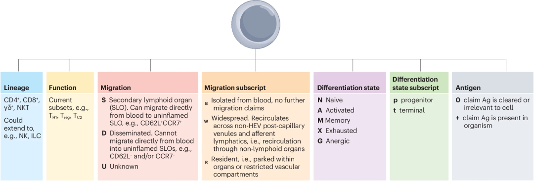 Fig 1, Masopust et al., 2025
Reference translation:
| Module | Options | Meaning |
|---|---|---|
| Lineage | CD4⁺, CD8⁺, γδ⁺ | Cell lineage |
| Migration | S | Lymph node-accessible |
| D | Lymph node-inaccessible | |
| U | Migration capacity unknown | |
| Migration subscript | B | Isolated from blood |
| R | Tissue-resident | |
| W | Widely circulating | |
| Differentiation | N | Naive |
| A | Activated | |
| M | Memory | |
| X | Exhausted | |
| G | Anergic | |
| Differentiation subscript | p | Progenitor |
| t | Terminal | |
| Antigen | 0 | Antigen cleared |
| + | Antigen persists |
Examples:
- CD8⁺ T
SM^0^ = Lymph node-accessible CD8⁺ memory T cell, antigen cleared (equivalent to classical TCM) - CD8⁺ T
DRM= Lymph node-inaccessible CD8⁺ tissue-resident memory T cell (equivalent to classical TRM) - CD8⁺ T
DXp^+^ = Lymph node-inaccessible CD8⁺ progenitor exhausted T cell, antigen persists (equivalent to TPEX)
The beauty of this system is that it never forces you to claim properties you haven't measured. If all you know is that the cells are CD62L⁻ CCR7⁻ and were isolated from blood, simply write CD8⁺ TDB — no need to agonize over whether they are TEM, TPM, or exTRM.
A Subset-Based Nomenclature Dictionary
In addition to proposing the new classification system, the paper also compiles existing T cell subsets with standardized definitions, serving as a convenient reference for researchers. Reproduced here:
Major Differentiation States
| State | Human | Mouse | Notes |
|---|---|---|---|
| Naive | CCR7^hi^, CD45RA⁺, CD45RO⁻, CD95⁻ | CD62L⁺, CD44^low^, CD11a^low^, CD122^low^ | No prior antigen stimulation |
| Activated | CD69⁺, CD25^hi^, KLF2^low^, Ki67⁺, HLA-DR⁺, CD38^hi^ | CD69⁺, CD25⁺, KLF2^low^, Ki67⁺ | Recently stimulated |
| Memory | CD11a^hi^, CD95⁺, CD58⁺, CD49d⁺ | CD44^hi^, CD11a^hi^, CD127⁺ | Long-lived, relatively quiescent |
| Anergic | Markers unclear | CD44⁺, FR4⁺, CD73⁺, FOXP3⁻, CD304⁺, CXCR5⁻ | Hyporeactive state |
| Virtual Memory | Markers unclear | CD44^hi^, CD49d^low^, CD62L^hi^, CD122^hi^ | Partial differentiation without antigen exposure |
CD8⁺ T Cell Subsets
| Subset | Human | Mouse | Notes |
|---|---|---|---|
| SLEC | CCR7⁻, CD45RA⁺, KLRG1⁺, TCF1^low^, T-bet^hi^, CX3CR1⁺, CD127^low^, CD27⁻, CD57⁺ | KLRG1⁺, CD127^low^ | Highly cytotoxic; unlikely to survive into memory phase |
| MPEC | Lacks universal markers | CD127⁺, CD27⁺, KLRG1^low^, TCF1⁺, T-bet^int^, CD62L^+/−^, CX3CR1⁻, CXCR3⁺ | High probability of differentiating into memory cells |
| EEC | KLRG1^low^, CD127⁻, CXCR3⁺, T-bet⁺, TCF1^low^, IFNγ⁺, TNF⁺ | KLRG1^low^, CD127⁻, CXCR3⁺, T-bet⁺, TCF1^low^, IFNγ⁺, TNF⁺ | Have not yet committed to SLEC or MPEC fate |
| LLEC | KLRG1^hi^, CD127^int^, CX3CR1^hi^, GZMB^hi^ | KLRG1^hi^, CD127^int^, CX3CR1^hi^, GZMB^hi^ | Long-lived but with poor proliferative capacity |
CD4⁺ T Cell Subsets
| Subset | Human | Mouse | Transcription Factor |
|---|---|---|---|
| T |
CXCR3⁺, CCR4⁻, CCR6⁻, CXCR5⁻, T-bet⁺, IFNγ⁺ | T-bet⁺, IFNγ⁺ | T-bet |
| T |
CRTh2⁺, CXCR3⁻, CCR4⁺, CCR6⁻, CXCR5⁻, GATA3⁺, IL-4⁺/IL-5⁺/IL-13⁺ | GATA3⁺, IL-4⁺/IL-5⁺/IL-13⁺ | GATA3 |
| T |
CXCR3⁻, CCR4⁺, CCR6⁻, CXCR5⁻, CCR8⁺, PU.1⁺, IL-9⁺, PPARγ⁺ (skin) | PU.1⁺, IL-9⁺ | PU.1 |
| T |
CXCR3⁻, CCR4^+/−^, CCR6⁺, CXCR5⁻, RORγt⁺, IL-17⁺ | RORγt⁺, IL-17⁺ | RORγt |
| CD4⁺ CTL | GZMB⁺, CCR7⁻, CD27⁻, may express CD95 | GZMB⁺, CCR7⁻, CD27⁻, may express CD95 | — |
| T |
CXCR5⁺, BCL6⁺ | CXCR5⁺, BCL6⁺ | BCL6 |
| GC T |
— | BCL6^hi^, CXCR5^hi^, PD-1^hi^, ICOS⁺, FR4^hi^, PSGL1^low^ | BCL6 |
| MZ T |
— | BCL6^low^, CXCR5^low^, PD-1^low^, FR4^low^, PSGL1^hi^ | BCL6 |
| T |
CXCR5⁺, BCL6⁺, CXCR4⁺, FOXP3⁻ | TCF1⁺, FR4⁺, P2RX7⁺, CXCR4⁺ | BCL6 |
| Treg | FOXP3⁺, CD25^hi^, CD127^low^, CTLA4^hi^, GITR^hi^ | FOXP3⁺, CD25^hi^, CD127^low^, CTLA4^hi^, GITR^hi^ | FOXP3 |
| tTreg | FOXP3⁺, CD25^hi^, CCR7^hi^, CD45RA⁺, CD45RO⁻ | FOXP3⁺, CD62L⁺, CD44^low^, Neuropilin-1^hi^, Helios^hi^ | FOXP3 |
| pTreg | FOXP3⁺, CCR7^hi^, CD45RA⁺, CD45RO⁻ | FOXP3⁺, CD62L⁺, CD44^low^, Neuropilin-1^low^, RORγt⁺ or GATA3^hi^, Helios^low/int^ | FOXP3 |
| eTreg | FOXP3^hi^, CD45RO⁺, CCR8^hi^, ICOS^hi^, IRF4⁺ | FOXP3^hi^, CD62L⁻, CD44^hi^, BLIMP1⁺, IL-10⁺, TGFβ⁺ | FOXP3 |
| T |
CXCR5⁺, FOXP3⁺, GITR⁺ | CXCR5⁺, PD-1⁺, BCL6⁺, FOXP3⁺, GITR⁺ | FOXP3, BCL6 |
Memory Subsets
| Subset | Human | Mouse | Notes |
|---|---|---|---|
| T |
CD62L⁺, CCR7⁺, CD27⁺, CX3CR1⁻, CD45RO⁺, CD45RA⁻ | CD44^hi^, CD62L⁺, CD122^hi^, TCF1⁺, CCR7⁺, CD103⁻, CX3CR1⁻ | Can circulate into lymph nodes |
| T |
CCR7⁻, CD45RA⁻, CD62L⁻ | CD44^hi^, CD62L⁻ | Cannot directly enter resting lymph nodes |
| T |
CCR7⁻, CD45RA⁺, CD27⁻, CX3CR1^hi^, CD57⁺ | — | Human-specific term |
| T |
KLF2^low^, CD69⁺, CD62L⁻, CCR7⁻, often CD103⁺, TCF1^low^, CD49a⁺, CD101⁺, CXCR6⁺ | KLF2^low^, CD69⁺, CD62L⁻, CCR7⁻, often P2RX7⁺, CD103⁺, TCF1^low^, CD49a⁺, CD101⁺, CXCR6⁺ | Tissue-resident; non-recirculating |
| ExT |
— | Phenotype resembles T |
Former T |
| T |
CD45RA⁻, CCR7^int^, CD27⁺ | CX3CR1^int^, CCR7^int^, CD27⁺ | Patrol between non-lymphoid tissues and blood |
| T |
CD95⁺, CCR7⁺, CD27⁺, CD28⁺, CD45RA⁺, Eomes⁻ | CD44^low^, CD62L⁺, CD122⁺, CD27⁺, Sca-1⁺, CD127⁺ | Stem cell-like; high self-renewal and differentiation potential |
Exhaustion-Related Subsets
| Subset | Key Markers | Notes |
|---|---|---|
| T |
PD-1⁺, TOX⁺, TCF1⁺, BCL6⁺, SLAMF6⁺, CXCR3⁺, LEF1⁺, CD28⁺, CD73⁺, XCL1⁺, CXCR5⁺, TIM3⁻, CD39⁻, CX3CR1^lo/int^, GZMB⁻ | Self-renewing; expands upon immune checkpoint blockade |
| T |
PD-1⁺, TOX⁺, TIM3⁺, T-bet⁺, GZMB⁺, Perforin⁺, IFNγ⁺, CX3CR1⁺, TCF1⁻, SLAMF6⁻, CD101⁻ | Retains effector function |
| T |
PD-1⁺, TOX⁺, TIM3⁺, GZMB⁺, CD39⁺, 2B4⁺, CD101⁺, TCF1⁻, SLAMF6⁻, CX3CR1⁻, CXCR3⁻ | Poor proliferative capacity; severely impaired function |
Innate-Like T Cells
| Subset | Human | Mouse |
|---|---|---|
| MAIT | CD3⁺, MR1-5-OP-RU tetramer⁺; Blood: CD3⁺, CD161⁺, Vα7.2⁺ | CD3⁺, TCRβ⁺, MR1-5-OP-RU tetramer⁺ |
| iNKT | CD3⁺, CD1d-αGalCer tetramer⁺; Blood: CD3⁺, Vα24Jα18⁺ or Vα24⁺/Vβ11⁺ | CD3⁺, TCRβ⁺, CD1d-αGalCer tetramer⁺ |
| γδ T (blood) | Predominantly Vγ9Vδ2⁺: CD161⁺, NKG2A⁺, CCR5⁺, CD16⁺ (post-activation); Vδ1⁺: TIGIT⁺, CD31⁺, CD127⁺, CD62L⁺, NKG2D⁺ | — |
| γδ T (intestine) | Predominantly Vδ1⁺: CD103⁺, CD38⁺, CD69⁺, CD101⁺, TIGIT⁺ | Vγ7⁺ IEL: TIGIT⁺, CD69⁺, CD122⁺, LAG3⁺, Thy1⁻, ~85% CD8αα⁺, Fcεr1γ⁺, CD3^hi^, CD5^low^ |
| γδ T (skin) | Predominantly Vδ1⁺: NKG2D⁺, CD69⁺, CXCR4⁺, CD44⁺, CD28⁻ | Epidermal Vγ5Vδ1⁺ DETC: CD45RB^hi^, CD3^hi^, CD103⁺; Dermal Vγ6Vδ1⁺: CD44⁺, CD69⁺, CD103⁺, CCR6⁺ |
| γδ T (spleen/LN) | — | γδIFNγ (Vγ1⁺): CD27⁺, CD44⁺, CCR6⁻; γδ17 (Vγ4⁺): CD44^hi^, CD27⁻, CCR6⁺, IL-17A⁺ |
| CD8αα⁺ TCRαβ | Most HLA-DR^hi^ | TL tetramer⁺, CD8β⁻ |
| DN T | TCR⁺, CD3⁺, CD4⁻, CD8αβ⁻, CD56⁻ | TCR⁺, CD3⁺, CD4⁻, CD8αβ⁻, NK1.1⁻ |
| Extrathymic DP | TCR⁺, CD3⁺, CD4⁺, CD8 (αα or αβ)⁺ | TCR⁺, CD3⁺, CD4⁺, CD8 (αα or αβ)⁺ |
Comparing the Two Classification Approaches
Despite the extensive work done to consolidate definitions across T cell subsets, the authors still advocate a swift transition to the new modular nomenclature. Here is a visual comparison of the two approaches:
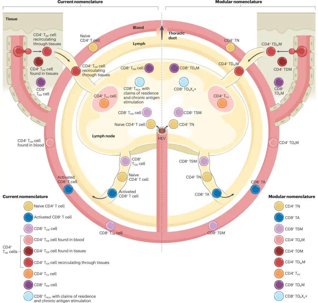 Fig 2, Masopust et al., 2025
...and how the same cell can be named under different hierarchical levels:
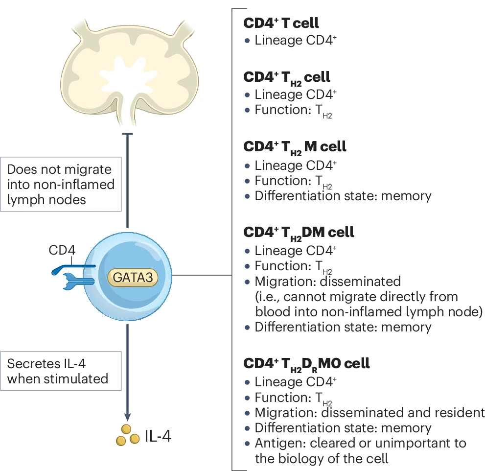 Fig 3, Masopust et al., 2025
Editor's Appendix: Recommended T Cell Annotation Tools
Standardized T cell nomenclature is unambiguously good news for bioinformaticians — it will greatly facilitate cross-dataset integration. Hierarchical, modular cell annotation is also increasingly the direction of travel for databases and computational tools alike.
Even before this guidelines paper was published, several groups had already been developing unified T cell annotation tools:
1. starCAT / TCAT (T-Cell Annotation Tool) (Nature Methods, 2025)
Released this year by the Raychaudhuri Lab (developers of Harmony), this rapid T cell annotation tool bypasses clustering entirely and instead quantifies 46 predefined gene expression programs (GEPs) covering core functional states including proliferation, cytotoxicity, exhaustion, and TH polarization. Available on GitHub and as a web app.
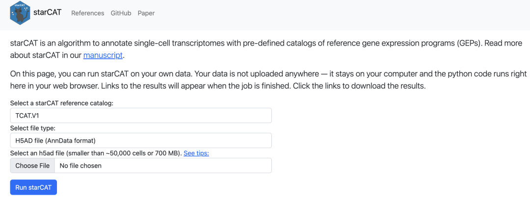
The web app runs entirely locally — no cloud dependency required.
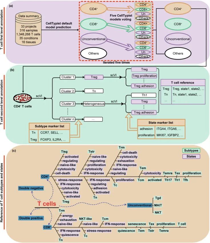
2. STCAT (Science Bulletin, 2025)
Builds the largest human T cell reference to date (1.34 million cells), organized into 33 subtypes and 68 subtype–state combinations. Uses a hierarchical model with marker-based correction. Available as a web app and on GitHub.
3. CellTypist (Science, 2022)
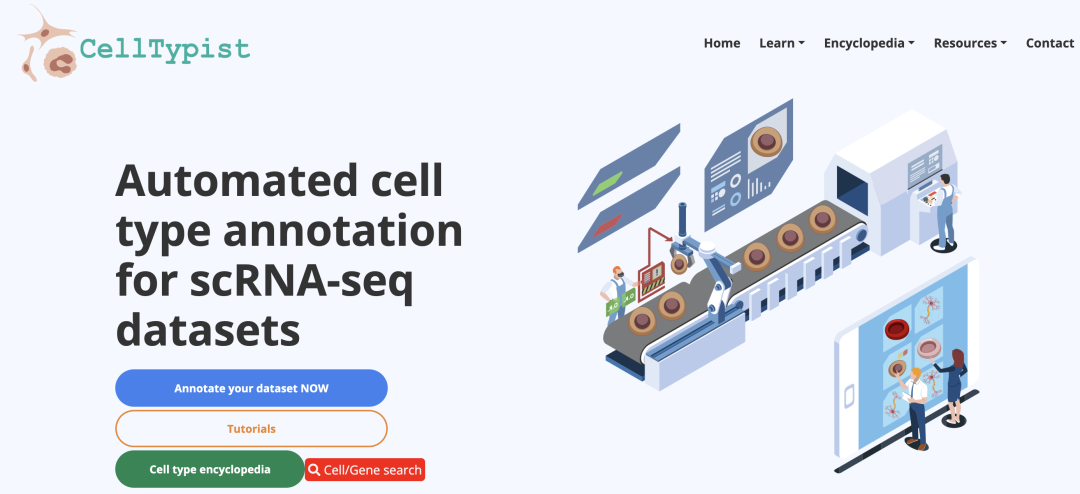
Available as a web app and a command-line tool on GitHub.
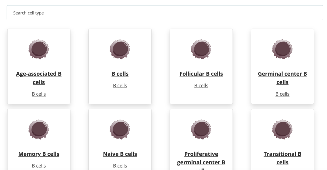
Developed by the Teichmann Lab, CellTypist is a fast logistic regression-based classifier that uses majority voting to improve prediction stability. Pre-trained models for immune cells are available out of the box for annotating new datasets. The web version also includes a "Cell Encyclopedia" for quickly looking up marker genes for any cell type.
4. CZ CELLxGENE (Nucleic Acids Research, 2025)
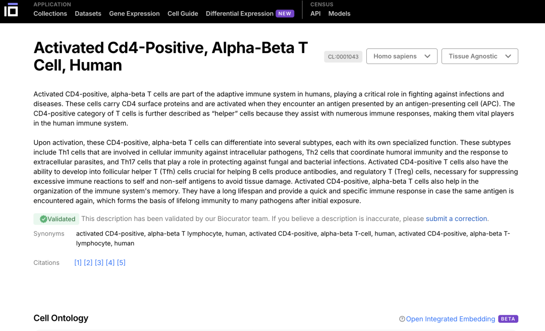
Web portal. This is far more than an annotation tool — it is a comprehensive data platform hosting over 93 million cells across 700+ cell types, with all datasets available for download in h5ad format. Its Cell Guide feature lets you look up detailed information on any cell type, including the cell lineage ontology and canonical marker genes. Extremely useful for reference and exploration.
References:
- Masopust D, et al. Guidelines for T cell nomenclature. Nat Rev Immunol. 2025. doi:10.1038/s41577-025-01238-2
- Kotliar D, et al. Reproducible single-cell annotation of programs underlying T cell subsets, activation states and functions. Nat Methods. 2025;22(9):1964–1980.
- Guo X, et al. An automatic annotation tool and reference database for T cell subtypes and states at single-cell resolution. Sci Bull. 2025.
- Domínguez Conde C, et al. Cross-tissue immune cell analysis reveals tissue-specific features in humans. Science. 2022;376(6594):eabl5197.
- CZI Cell Science Program, et al. CZ CELLxGENE Discover: a single-cell data platform for scalable exploration, analysis and modeling of aggregated data. Nucleic Acids Res. 2025;53(D1):D886–D900. doi:10.1093/nar/gkae1142
Originally posted on WeChat Official Account: bioinfo_studyroom