Rahul Satija's Eight Major Single-Cell Breakthroughs of 2025


2025Eight Major Single-Cell Breakthroughs
2025 Single Cell Genomics Day
On April 25, 2025, the Satija Lab at New York University — home to the developers of the Seurat package — hosted the 9th annual Single Cell Genomics Day online. Dr. Rahul Satija presented eight exciting new advances spanning both experimental and computational innovations. Let's dive in!
1. Combinatorial Droplet-Based Barcoding
A major limitation of conventional 10x Genomics technology is that the vast majority of droplets are empty, leading to wasted reagents and higher costs. New approaches such as UDA-seq overcome this with a clever combinatorial barcoding strategy:
- A large number of cells are loaded into the 10x instrument so that multiple cells enter the same droplet
- Cells remain intact and are not lysed during this step
- Cells are then transferred to a 96-well plate where a second barcode is added
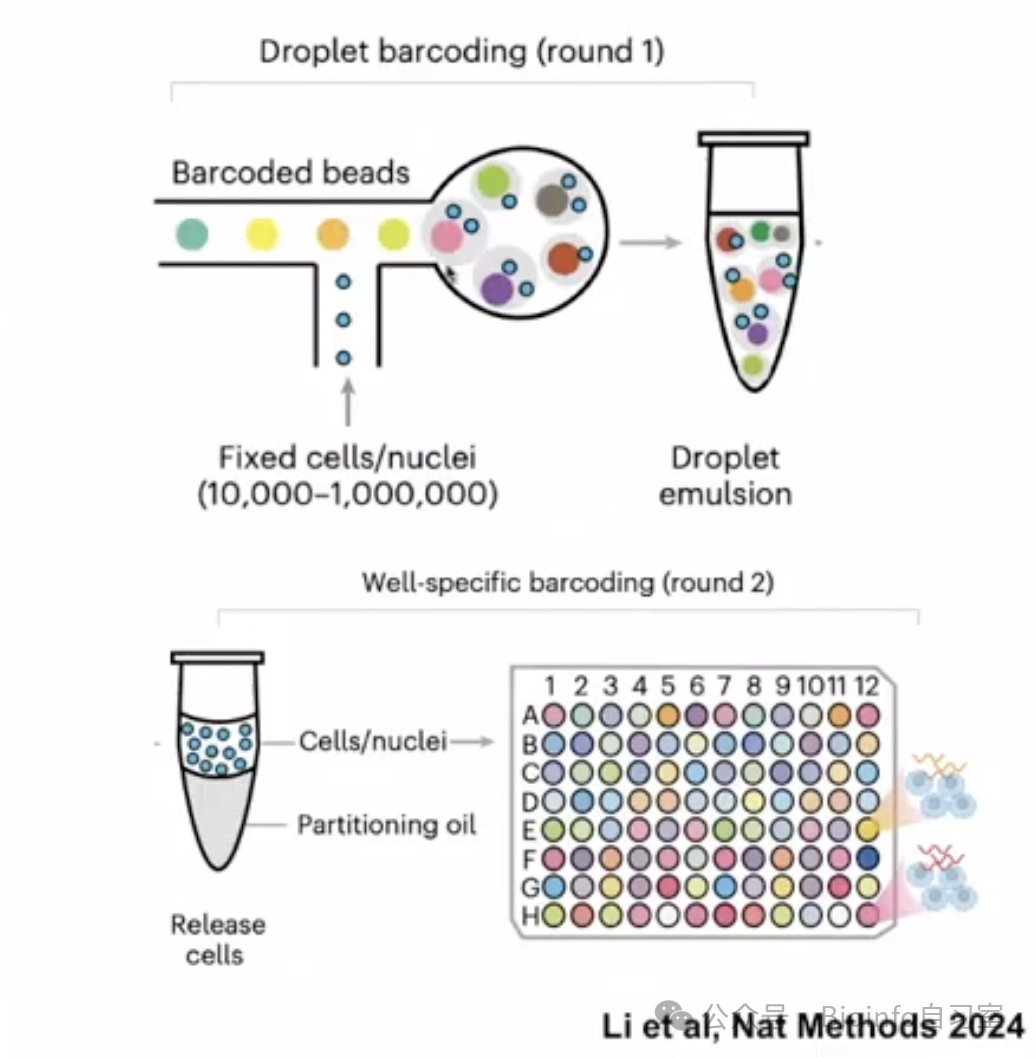
Combinatorial barcoding strategy (reference cited in figure)
This approach offers substantial advantages:
- Approximately 150,000 cells can be profiled in a single run (vs. 5,000–10,000 with conventional methods)
- Dramatically lower cost (~90% reduction)
- Compatible with RNA-seq, ATAC-seq, and multiomics assays
- Validated in human blood, retina, kidney, and other tissues
2. Organismal-Scale Reference Mapping
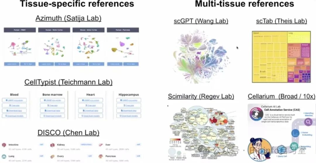
Dr. Satija's overview of existing reference annotation strategies
As single-cell datasets grow exponentially, reference mapping has become increasingly important. Just as bulk RNA-seq reads are aligned to a reference genome to facilitate interpretation, researchers apply the same logic to scRNA-seq. Reference mapping tools generally fall into two categories: tissue-specific references and whole-body references (e.g., foundation models such as scGPT). However, these models often suffer from inconsistent annotation across training datasets.
The PanHuman Azimuth neural network, developed by the Satija Lab, addresses this with a unified cell ontology that can:
- Annotate cells consistently across the entire human body
- Generalize across tissue types and sequencing technologies
- Leverage carefully curated training data from 23 tissues
- Apply a hierarchical, standardized cell classification scheme
The tool has successfully mapped the Tabula Sapiens V2 reference dataset and approximately 85 million human cells from the ARC Institute — all in under a day. PanHuman Azimuth is open-source, supports both R and Python, and is compatible with Seurat and Scanpy.

PanHuman Azimuth usage guide — give it a try!
3. Spatial Analysis without Imaging
Traditional spatial transcriptomics platforms (e.g., Slide-seq, Slide-tags) require elaborate puck sequencing and microscopy imaging steps to determine the spatial coordinates of barcoded beads. What if you could recover spatial positions without any imaging at all?
Researchers exploited a clever mathematical trick: reconstructing spatial positions from pairwise distances between beads — much like reconstructing a map of the United States from a table of inter-city distances. The key innovation is the introduction of "diffusible beads" that act as random "broadcasters." If two beads capture signal from the same broadcaster, they must be spatially proximate.
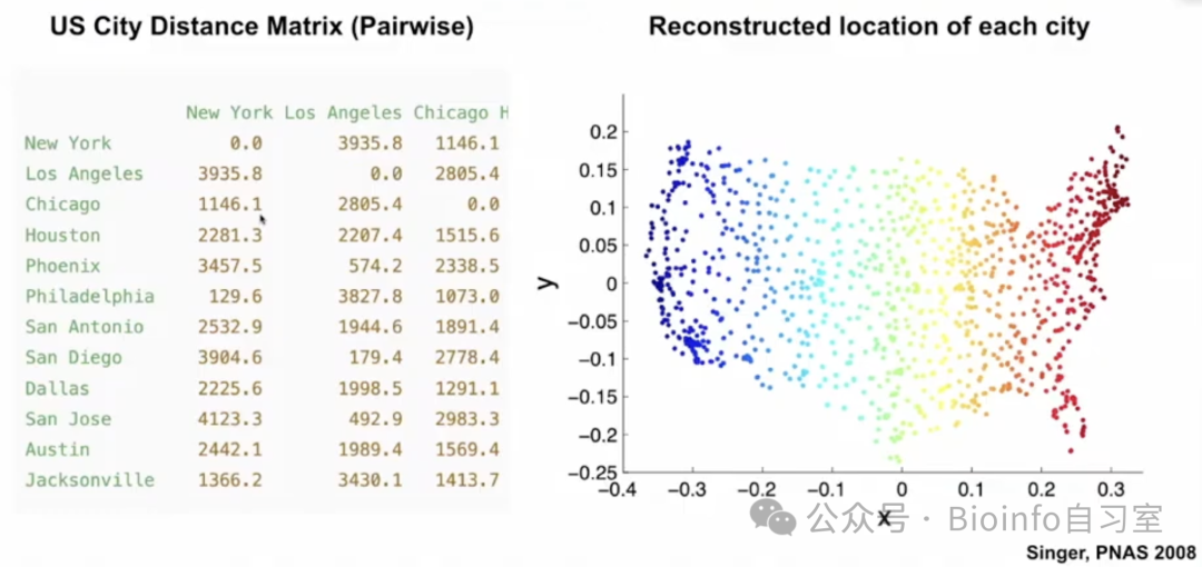
Illustration: reconstructing a U.S. map from pairwise city distances (reference cited in figure)
This approach has been validated in mouse brain and intact P1 head samples. By dramatically reducing the cost and complexity of acquiring spatial information, it promises to accelerate the convergence of single-cell and spatial genomics.
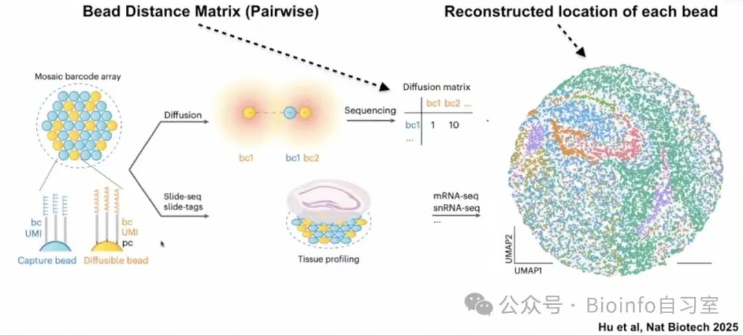
Diffusion matrices derived from "broadcaster beads" enable 3D reconstruction of tissue architecture (reference cited in figure)
4. Semi-Permeable Droplet Systems
Conventional droplet-based systems are sealed after encapsulation, making it impossible to add or remove reagents — a limitation that restricts experimental complexity. CAGE (Capsule with gel envelope) technology breaks this barrier with a hydrogel shell made of F127DA polymer featuring 10–15 nm pores:
- Small molecules (enzymes, short DNA primers, culture media) can freely diffuse in and out
- Larger molecules (cDNA, long nucleic acids) are retained inside the capsule
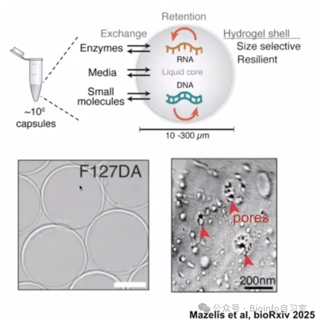
Schematic of hydrogel capsules (reference cited in figure)
This enables:
- Multi-step reactions and wash steps at single-cell resolution
- Cell growth within capsules to generate small clonal populations
- Differential perturbation treatments across capsules, followed by molecular readouts
CAGE opens exciting new possibilities for single-cell experiments, particularly those requiring long-term culture and complex multi-step processing. Preprints from the Klein Lab and others are available on bioRxiv, and Atroni Biosciences is commercializing the technology.
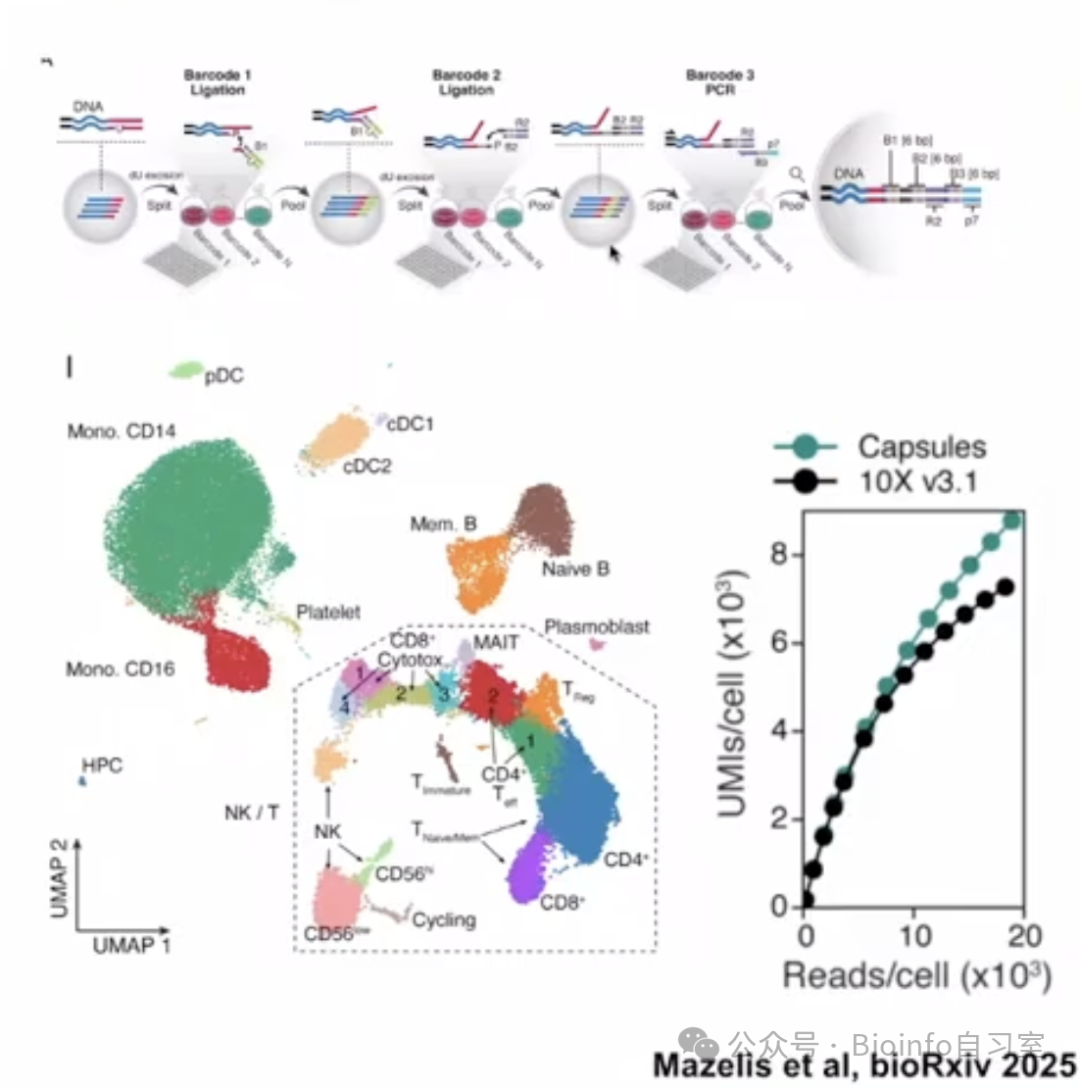
Example: single-cell sequencing performed within CAGE capsules
5. Sample-Level Analysis of Single-Cell Data
Single-cell analysis has traditionally focused on characterizing individual cells and cell types. Multiple groups have recognized, however, that in case-control studies, analyzing samples as units is often more clinically meaningful. This is a key frontier: how do we move beyond cell-level analysis to sample-level analysis?
Three key directions for sample-level analysis:
- Clustering: Cell clustering reveals cell types; sample clustering may reveal patient subgroups or prognostic stratification
- Trajectories: Cell trajectories represent differentiation; sample trajectories may capture disease progression or severity
- Reference mapping: In addition to annotating cell types, reference mapping can be extended to predict patient clinical outcomes or treatment responses
scPoli is a variational autoencoder framework for sample-level analysis that enables PCA at the sample level rather than the cell level, allowing visualization of how different individuals group based on their cellular composition.
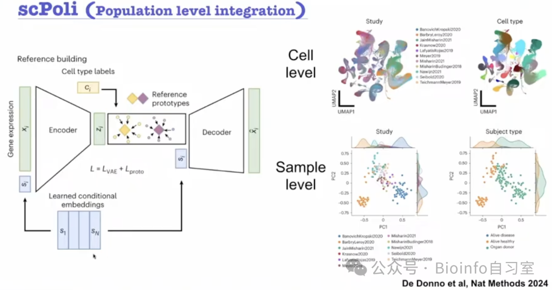
scPoli schematic: clustering and integration at the sample/dataset level (reference cited in figure)
Another highly influential method is scLKME, which shifts focus from individual cells to the overall density of cell-state distributions within a sample. In a study of approximately 40 neonatal cord blood samples, scLKME learned sample-level trajectories with pseudotime strongly correlated with gestational age — offering a new perspective on how the density of immune cell states in the neonatal immune system changes over developmental time.
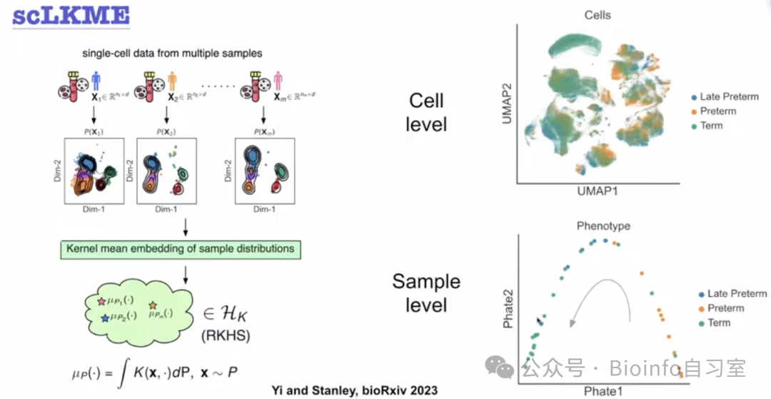
Individual-level pseudotime for studying developmental trajectories (reference cited in figure)
6. Native Cell Lineage Tracing
Reconstructing a complete organismal lineage tree is a major challenge: somatic mutation rates are low, making it difficult to infer lineage relationships from DNA sequence alone. Traditional approaches have relied on either lentiviral integration-based clonal barcoding (e.g., CellTag) or mitochondrial DNA mutations, but each has limitations and neither scales robustly to primary human tissues.
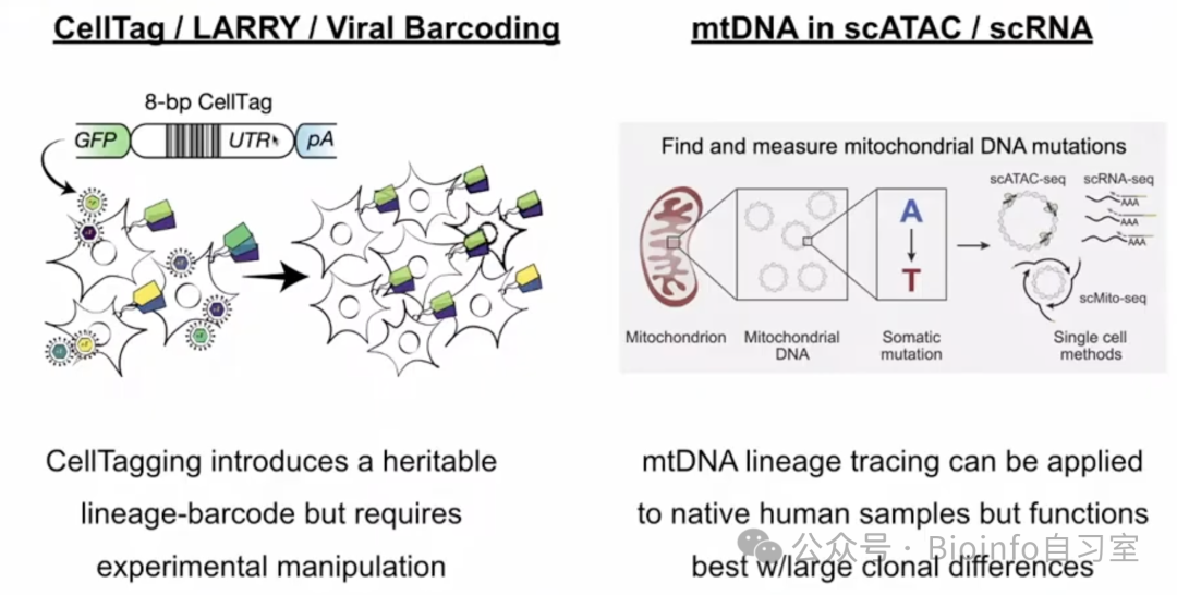
Overview of traditional lineage tracing strategies
A key breakthrough this year exploits epimutations — stochastic changes in epigenetic state — for lineage reconstruction. DNA cytosine residues can acquire methylation marks that are heritable through cell division, but with approximately a 0.1% probability of changing at each division. If two cells have nearly identical DNA methylation profiles, they are likely clonally related — possibly even sister cells.
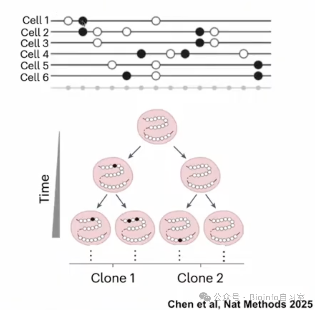
Epimutation-based lineage tracing approach (reference cited in figure)
Researchers at Westlake University developed MethylTree, demonstrating in a benchmark experiment with serially passaged and split HEK cells that clustering based on DNA methylation profiles yields clusters that align remarkably well with known lineage structure. The Velten Lab pursued a similar strategy in mammalian hematopoietic stem cells, revealing clonal substructure when clustering on neutral CpG sites.
7. Building Virtual Cells with AI
A virtual cell is a computational model capable of simulating and predicting a cell's past, present, and future behavior. This concept has swept through systems biology this year. While the idea of digitally modeling cells is not new, we now have the data generation methods (single-cell technologies) and analytical tools (AI) to make it a reality.
The goal is to run in silico experiments and predict outcomes — for example, how a cell will respond to a drug or genetic perturbation. This won't fully replace wet-lab experiments, but it can dramatically improve experimental design. If you have 50,000 candidate compounds that cannot all be tested experimentally, virtual screening can prioritize the most promising 1,000 for follow-up.
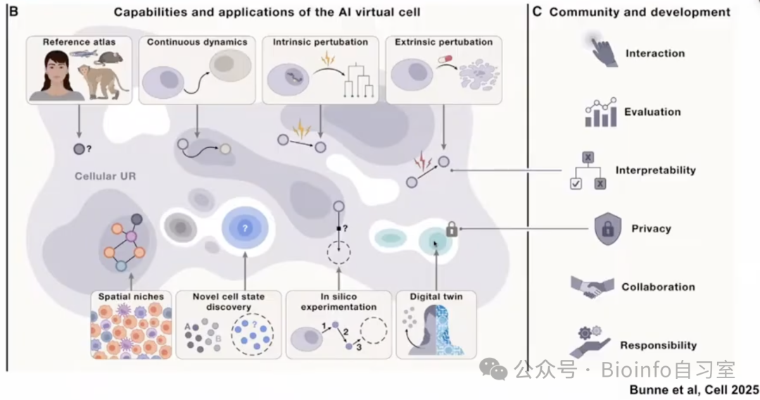
Applications of AI-based virtual cells (reference cited in figure)
Prophet, developed by the Fabian Theis Lab, uses a Transformer architecture trained on diverse single-cell and perturbation datasets. It aims to scalably and universally predict cellular phenotypes including cell viability, gene expression, and morphology. As a demonstration, they performed virtual EGFR inhibitor screening across a large panel of lung cancer cell lines, predicting sensitivity gradients that aligned with experimental results. While these predictions still require high-throughput validation, the broader vision is to use virtual cells as an initial filter to prioritize candidate cell lines, drug targets, and therapies.
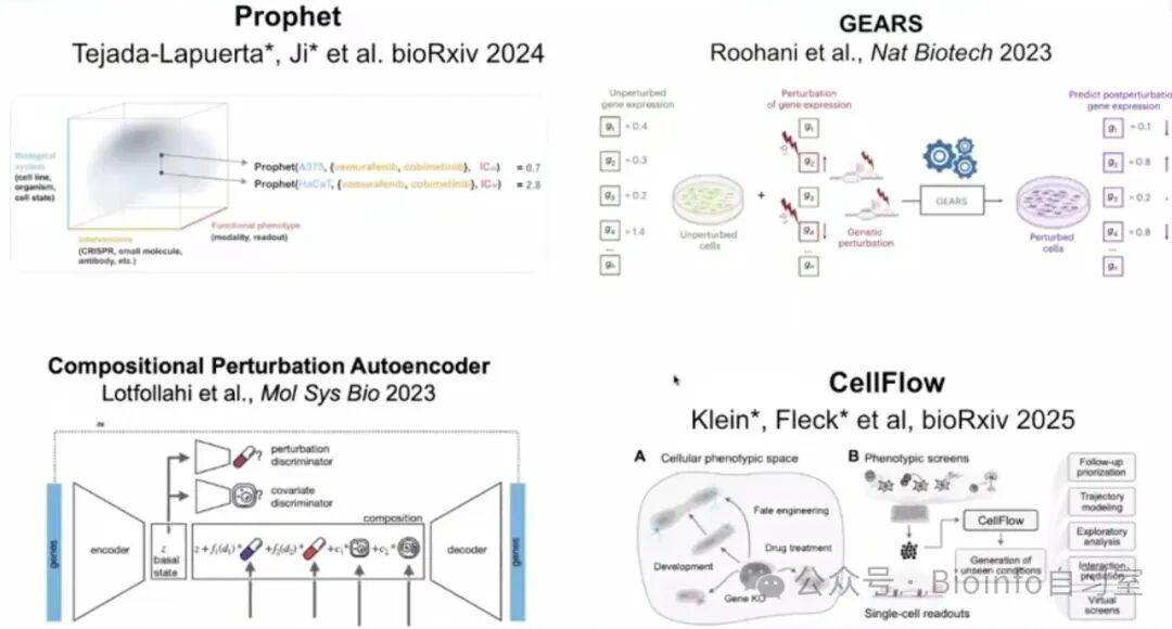
Selected virtual cell frameworks (reference cited in figure)
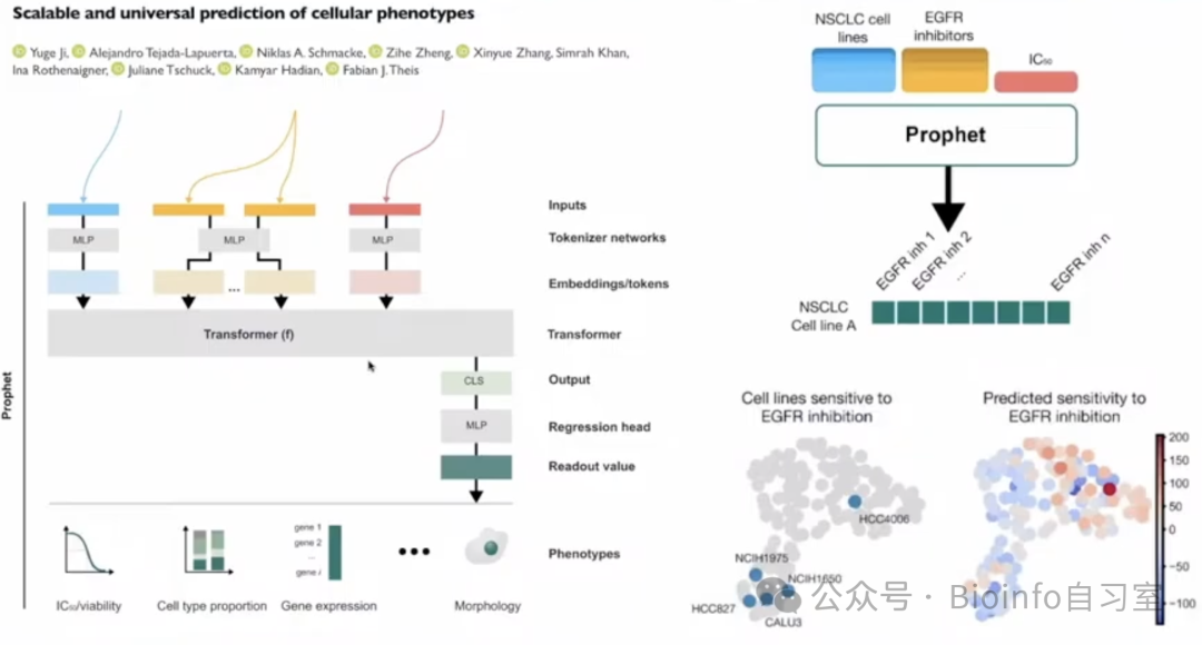
The Prophet framework (reference cited in figure)
8. Inferring Transcription Factor Binding from Single-Cell Data
Inferring transcription factor (TF) binding and its dynamics in single cells has been an extreme challenge: TF–DNA interactions are inherently weak and transient. Recently, the Buenrostro Lab (one of the groups that invented single-cell ATAC-seq) discovered that TF occupancy creates a local decrease in chromatin accessibility — a "footprint" — at binding sites. Building on this, they developed seq2PRINT, a deep learning model that integrates DNA sequence information with ATAC-seq signal to enhance footprint detection and track TF dynamics during differentiation, or to directly predict TF binding.
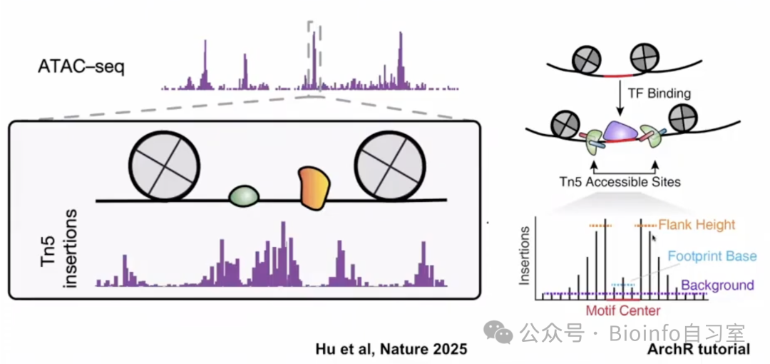
TF binding sites display local reductions in chromatin accessibility — the "footprint" (reference cited in figure)
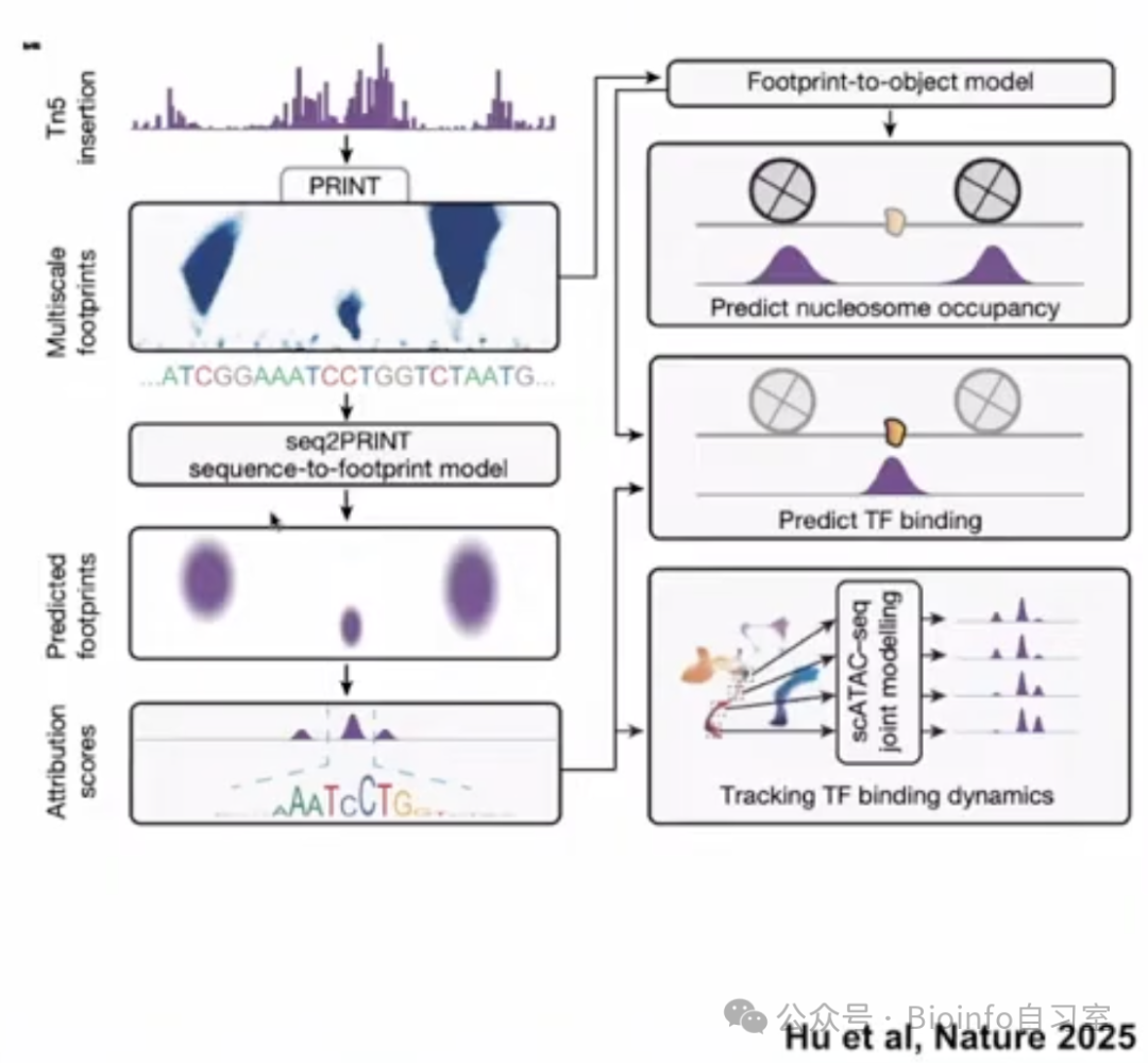
seq2PRINT tracks TF dynamics through multi-model inference (reference cited in figure)
A complementary experimental strategy — D&D-seq (docking and deamination) — was developed by the Raimondi and Landau Labs. This approach uses a secondary nanobody fusion protein to recruit a potent base editor to TF-occupied genomic loci. The base editor introduces permanent C→U mutations at those sites, which can then be detected in single-cell ATAC readouts. The main limitation is data sparsity: fewer than 100 binding events are captured per cell, necessitating deep sequencing of large cell numbers and pseudobulk aggregation by cell type. As sequencing costs continue to decline and large-scale experiments become more practical, these computational and experimental approaches will bring us steadily closer to the ultimate goal: resolving TF binding dynamics at single-cell resolution.
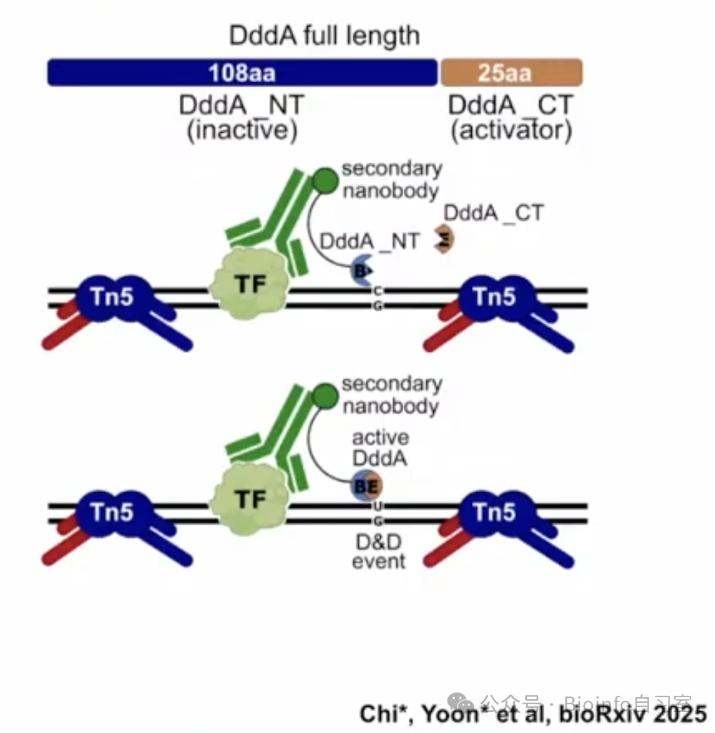
D&D-seq permanently marks TF binding events through base editing (reference cited in figure)
-END-
Follow Bioinfospace for updates on single-cell frontiers, in-depth single-cell tutorials, and bioinformatics tips! Star ✨ us to never miss a post:
Originally posted on WeChat Official Account: bioinfo_studyroom