Paper Outlook — September 14-20, 2026

TL;DR
- Single-cell and spatial mapping dominates the week, with new atlases of the human adrenal gland, the postnatal mouse heart, and the developing human uterus showing how cell-type-resolved maps are now standard for tissue biology.
- Two cross-species comparisons (human vs. mouse adrenal glands, dog vs. human bone cancer) point to both conserved biology and clear limits of animal models, a useful reminder for anyone designing translational studies.
- Two mutation-focused papers link environment to genome-level outcomes: caloric restriction reduces DNA damage accumulation across mouse tissues, while atypical UV lesions in single-stranded DNA help explain unusual driver mutations in melanoma.
- Functional genomics is moving in vivo, exemplified by spatially guided knockdown of gene regulators in living mouse hearts to test roles in maturation.
- The unifying thread is the integration of cell-by-cell data with spatial context and functional validation, which together reveal biology that bulk methods simply miss.
Human and mouse adrenal glands are characterized by species-specific steroidogenic states and tissue turnover.
Scientists built detailed cellular maps of the adrenal gland, the organ that produces stress hormones, in both adult humans and mice. Humans turn out to have unique cell types that can directly make cortisol, along with structural features, such as a distinct inner zone, that simply do not exist in mice. Because mice are commonly used as stand-ins for human adrenal biology, these differences raise a practical question: how much of what we learn in mice actually carries over to human disease and drug testing?
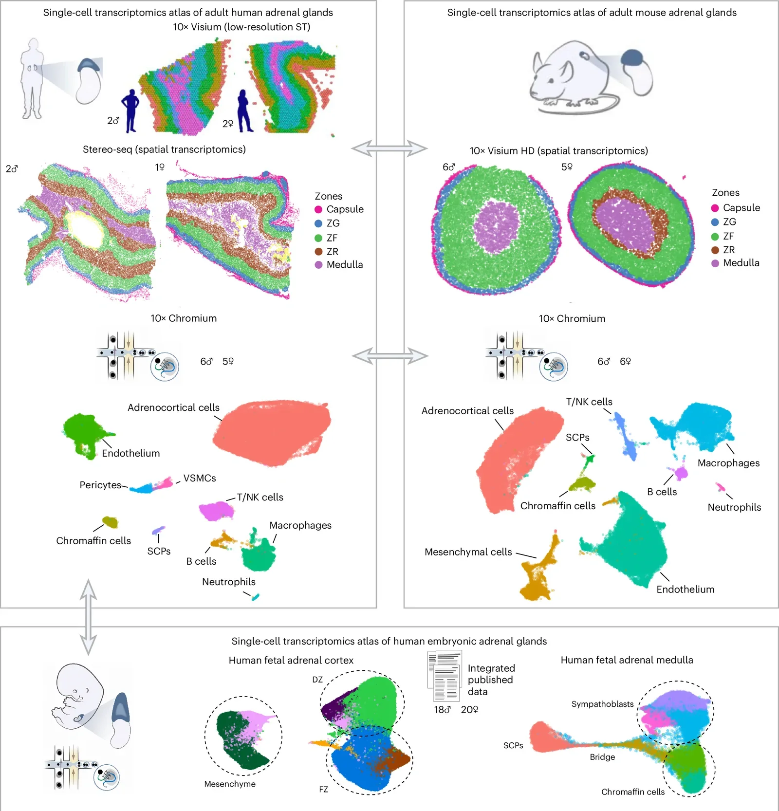
Fig. 1 from Kastriti ME et al., Nature genetics (2026). Licensed CC BY. Source: 10.1038/s41588-026-02737-1.
Nature genetics (2026)
Read the original → https://doi.org/10.1038/s41588-026-02737-1
Integrated Transcriptomics of Human and Canine Osteosarcoma Reveals Species Stemness and Trabectedin Sensitivity.
Researchers compared bone cancer in dogs and humans at the molecular level to ask when studying dogs can reliably predict how human patients will respond to treatment. They found shared features alongside species-specific differences in how tumors behave and react to the drug trabectedin. The practical upshot is a clearer view of which canine cancer studies are likely to translate to humans, and which are more likely to mislead.
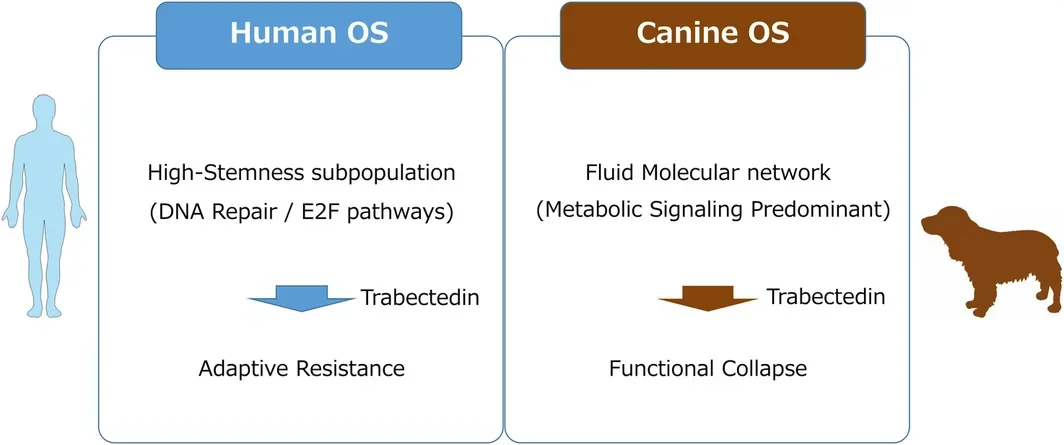
FIGURE 6 from Kono K et al., Cancer science (2026). Licensed CC BY-NC-ND. Source: 10.1111/cas.70524.
Cancer science (2026)
Read the original → https://doi.org/10.1111/cas.70524
Caloric restriction modulates genome-wide somatic mutation in mice.
Mice placed on a calorie-restricted diet accumulated fewer DNA mutations in their liver, kidney, and brain cells compared with mice eating normally, with the strongest effect on a common age-related mutation pattern. That ties the well-known lifespan-extending effect of caloric restriction to a concrete decrease in DNA damage at the genome level, adding a mechanistic angle to why eating less may slow aspects of aging.

Cell (2026)
Read the original → https://doi.org/10.1016/j.cell.2026.08.013
Widespread atypical UV-induced mutations form in single-stranded DNA.
UV light typically damages DNA through well-known lesions that produce classic C-to-T mutation patterns in skin cancers. This study highlights an underappreciated pathway: unusual UV damage that arises when DNA is briefly single-stranded during copying or repair, generating atypical mutations seen in key melanoma driver genes. It broadens how scientists think about UV-induced skin cancer and why some driver mutations have been hard to explain.
Science advances (2026)
Read the original → https://doi.org/10.1126/sciadv.aeg5184
Spatially guided in vivo single-cell functional genomics of postnatal heart.
Researchers combined single-cell gene activity measurements with spatial location data to build a detailed molecular map of how the mouse heart matures after birth. They used this map to nominate key gene regulators of heart cell maturation and then tested their function directly in living mice. The resulting atlas is released as a public resource for studying heart development, congenital disease, and regeneration.
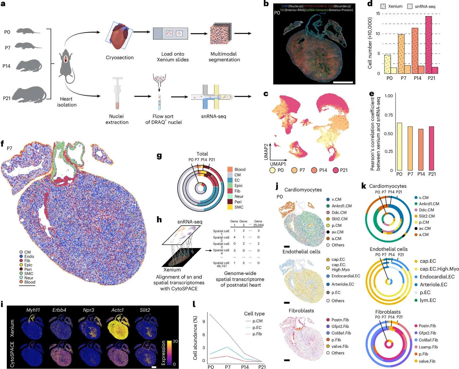
Fig. 1 from Wang H et al., Nature cardiovascular research (2026). Licensed CC BY-NC-ND. Source: 10.1038/s44161-026-00861-z.
Nature cardiovascular research (2026)
Read the original → https://doi.org/10.1038/s44161-026-00861-z
HMMR<sup>+</sup> stromal cells drive human uterine development.
A study used single-cell gene activity and spatial mapping to trace how the human uterus forms before birth, from the early second trimester through adolescence. The authors identified a specific cell population, marked by the HMMR gene, that is highly proliferative and gives rise to both the uterine lining and muscle layers. The work lays a foundation for understanding reproductive disorders and the developmental origins of uterine disease.
Cell discovery (2026)
Read the original → https://doi.org/10.1038/s41421-026-00921-5
Quick links
| Paper | Venue / Year | Link |
|---|---|---|
| Human and mouse adrenal glands are characterized by species-specific steroidogenic states and tissue turnover. | Nature genetics (2026) | Read → |
| Integrated Transcriptomics of Human and Canine Osteosarcoma Reveals Species Stemness and Trabectedin Sensitivity. | Cancer science (2026) | Read → |
| Caloric restriction modulates genome-wide somatic mutation in mice. | Cell (2026) | Read → |
| Widespread atypical UV-induced mutations form in single-stranded DNA. | Science advances (2026) | Read → |
| Spatially guided in vivo single-cell functional genomics of postnatal heart. | Nature cardiovascular research (2026) | Read → |
| HMMR<sup>+</sup> stromal cells drive human uterine development. | Cell discovery (2026) | Read → |