Learning Single-Cell Analysis with Theis Lab: An Overview of Single-Cell Sequencing

Cracking single-cell analysis with Theis Lab
Series No.: 02 | Published: 2024-12-13 | Estimated reading time: 15 minutes
Single-Cell Best Practices02. Single-Cell Sequencing⭐️ Chapter Overview:
- A brief introduction to cell theory
- A brief history of first-, second-, and third-generation sequencing
- The principles of RNA sequencing
- Leading approaches in single-cell sequencing
2.1 The Basic Unit of Life
The cell is the fundamental unit of all living organisms. In a sense, a cell is like a miniature precision factory — one that can maintain homeostasis, carry out metabolism, grow, adapt to its environment, reproduce, respond to stimuli, and self-organize. This "little factory" was first observed in 1665 by the British scientist Robert Hooke, who peered at a thin slice of cork through a rudimentary microscope and noticed it resembled a honeycomb. He named those tiny compartments "cells."
In 1839, Matthias Jakob Schleiden and Theodor Schwann formally proposed cell theory, which states: all living organisms are composed of cells; the cell is the basic unit of life; and all cells arise from pre-existing cells.
Since cell theory was established, scientists have made further discoveries:
- Energy flows within cells
- Genetic information is transmitted between cells in the form of DNA
- The chemical composition of all cells is essentially similar
Cells fall into two main types: eukaryotic cells and prokaryotic cells.
- Eukaryotic cells have a nucleus enclosed by a nuclear membrane, which houses the chromosomes
- Prokaryotic cells have only a nucleoid region and lack a true nucleus
The etymology of "eukaryote" is quite interesting: nucleus comes from the Latin word for "kernel" or "seed." Eukaryotes can be either unicellular or multicellular, whereas all prokaryotes are unicellular. Another hallmark of eukaryotic cells is the presence of numerous membrane-bound organelles, each performing a specific function.
Compared to prokaryotic cells, eukaryotic cells are approximately 10,000 times larger. They possess a rich internal architecture, including various organelles and a cytoskeleton built from microtubules, microfilaments, and intermediate filaments. The DNA replication machinery within the nucleus reads and copies genetic information, sustaining the cell cycle.
In eukaryotes, DNA is organized into multiple linear chromosomes, which are separated by mitotic spindles during cell division. Understanding the genetic information encoded in DNA is essential for studying evolution and disease-related processes. Sequencing is the process of decoding the nucleotide sequence of DNA, and it can be used to:
- Identify the locations and functions of genes and regulatory elements within DNA molecules and genomes
- Discover genomic features such as open reading frames (ORFs) or CpG islands (which typically mark promoter regions)
- Compare DNA sequences across species to study evolutionary relationships
- Investigate associations between mutations and disease or disease resistance
As an example, sickle cell disease is a group of red blood cell disorders caused by abnormal hemoglobin — the protein responsible for carrying oxygen. This leads to a range of serious health complications, including:
- Pain crises
- Anemia
- Swelling of the hands and feet
- Bacterial infections
- Stroke
The disease arises when a person inherits one defective copy of the β-globin gene (HBB) from each parent. Specifically, a GAG codon in the β-globin gene is changed to GTG, causing the sixth amino acid to switch from glutamic acid to valine — the E6V mutation — triggering the disease.
It is worth noting, however, that not all diseases can be traced to such a straightforward single-nucleotide variant. Most diseases arise from far more complex regulatory processes.
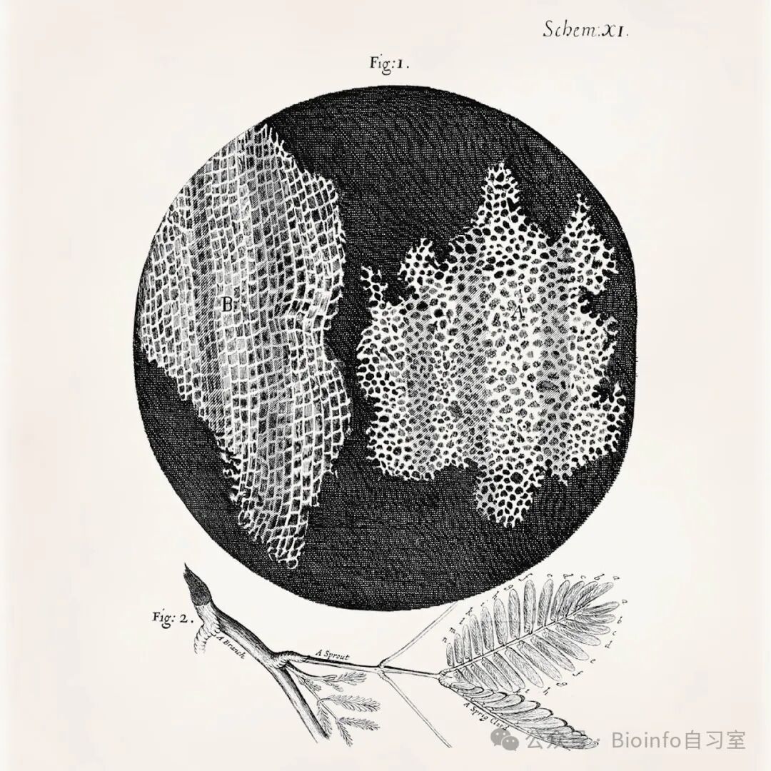
Figure 2.1: Robert Hooke's illustration of cork cells. Image from Micrographia.
2.2 A Brief History of Sequencing Technology
2.2.1 First-Generation Sequencing
Although DNA was first isolated by Friedrich Miescher as early as 1869, the scientific community took over a century to develop high-throughput sequencing technologies. Some important milestones include:
- 1953: Watson, Crick, and Franklin discover the double-helix structure of DNA
- 1965: Robert Holley sequences the first tRNA
- 1972: Walter Fiers sequences the first complete gene (the coat protein of bacteriophage MS2)
- 1970s: Sanger develops the "chain termination method" (commonly known as "Sanger sequencing")
- 1987: Leroy Hood and Michael Hunkapiller develop the ABI 370 instrument, automating Sanger sequencing
The main advantages and disadvantages of Sanger sequencing are as follows:
Advantages
- Simple to operate; moderately priced
- Very high accuracy (error rate < 0.001%)
Disadvantages
- Can only sequence short fragments of 300–1,000 bases
- The first 15–40 bases are typically of lower quality (due to primer-binding)
- Sequence quality drops off after 700–900 bases
- If the DNA fragment was cloned, vector sequences may contaminate the read
- Per-base cost is higher than second- or third-generation sequencing
2.2.2 Second-Generation Sequencing
In 1996, Mostafa Ronaghi, Mathias Uhlen, and Pål Nyren introduced a new DNA sequencing technology — pyrosequencing — marking the dawn of the second-generation sequencing era. Next-generation sequencing (NGS) became possible thanks to three key advances:
- Laboratory automation
- Computational power
- Miniaturization of reactions
Pyrosequencing works by measuring the luminescent signal generated during the sequencing reaction, a process also known as "sequencing-by-synthesis."
Two years later, Shankar Balasubramanian and David Klenerman at Solexa improved upon this approach, developing a new method using fluorescent dyes. This technology became the foundation of Illumina sequencing instruments and remains the dominant platform on the market today.
Other important milestones include:
- 2005: The Roche 454 sequencer debuts, fully automating the pyrosequencing process for the first time
- 2007: The SOLiD system introduces "sequencing-by-ligation" technology
- 2011: Life Technologies launches Ion Torrent, which uses sequencing-by-synthesis to detect hydrogen ions released during DNA synthesis
Second-generation sequencing remains widely used today. Its advantages and disadvantages are:
Advantages
- Relatively low reagent costs
- Can work with scarce sample material
- High sensitivity for detecting low-frequency variants
- Comprehensive genome coverage
- High throughput; multiple samples can be run in parallel
- Thousands of genes can be sequenced simultaneously
Disadvantages
- Instruments are expensive and often shared among multiple users
- Large instrument footprint; not suited for fieldwork
- Short reads (reads) are disadvantageous for de novo genome assembly
- Sequencing quality depends on the availability of a reference genome
2.2.3 Third-Generation Sequencing
Third-generation sequencing (sometimes also called "next-next-generation sequencing") brought two major innovations:
- Long-read sequencing: Compared to Illumina's short reads (75–300 bp), third-generation platforms generate much longer nucleotide fragments. This is particularly important for species that lack a reference genome.
- Real-time sequencing: Combined with portable instruments, sequencing is no longer confined to the laboratory — it can now be performed in the field.
Key technologies:
PacBio sequencing (2010):
- Uses nanopore technology (SMRT cells)
- Typical read lengths of 8–15 kb, with a maximum exceeding 70 kb
- Allows direct observation of the incorporation of individual nucleotides
Oxford Nanopore sequencing (2012):
- Introduced portable sequencers: GridION and its successors MinION and Flongle
- Read lengths can exceed 2 Mb
- Instruments small enough to fit in the palm of your hand
- Identifies sequences by monitoring changes in electrical current as nucleic acids pass through protein nanopores
Third-generation sequencing addresses the long-read shortcomings of NGS, though it comes with its own trade-offs:
Advantages
- Long reads facilitate the assembly of large, novel genomes
- Portable instruments suited for fieldwork
- Can directly detect epigenetic modifications on DNA and RNA
- Fast sequencing speed
Disadvantages
- Higher error rates than second-generation sequencing
- Reagent costs are generally higher than second-generation sequencing
2.3 Overview of the NGS Workflow
Despite the diversity of NGS technologies, the fundamental steps for sequencing DNA (and RNA after reverse transcription) are largely the same. The key differences lie in the sequencing chemistry used by each platform.
Three main steps in sequencing:
Sample preparation and library construction:
- The DNA sample is first fragmented into smaller pieces
- Adapter molecules are ligated to the fragments; these adapters serve two purposes:
- Help the DNA fragments bind to the sequencing flow cell or chip
- Provide primer-binding sites
Amplification and sequencing:
- The library is converted into single-stranded molecules
- An amplification step (e.g., PCR or bridge amplification) generates clusters of DNA molecules
- During a sequencing run, all clusters undergo independent reactions simultaneously
Data output and analysis:
- Different sequencers produce different output formats:
- Some generate fluorescent signals
- Others generate electrical signals
- Raw data volumes are typically very large
- Complex computational processing is required
- Different sequencers produce different output formats:
2.4 RNA Sequencing
Up until now, we have been talking mostly about DNA sequencing. But knowing the DNA sequence and the locations of regulatory elements alone cannot tell us about the real-time dynamics of a cell. For example, a single gene can produce multiple proteins through alternative mRNA splicing, and splice variants can render an enzyme non-functional, leading to disease. This is precisely why we need RNA sequencing (RNA-Seq).
The basic workflow of RNA-Seq is similar to that of DNA sequencing, but it requires an additional reverse transcription step to convert RNA into complementary DNA (cDNA). RNA-Seq gives researchers a "snapshot" of the gene expression profile — the transcriptome — of a cell, tissue, or organism at the moment of sequencing. This information can be used to detect changes in gene expression across a wide range of conditions, such as disease states, drug responses, or different environmental contexts.
Compared with traditional methods of measuring gene expression (such as microarrays and RT-qPCR), RNA-Seq has one major advantage: it requires no prior probe design. This means RNA-Seq can unbiasedly sample transcripts, enabling the discovery of:
- Novel gene isoforms
- Gene fusions
- Single-nucleotide variants
- Other unexpected transcriptional features
This freedom from prior assumptions makes RNA-Seq datasets particularly well-suited for exploratory analysis, helping researchers uncover new biological phenomena.
2.5 Single-Cell RNA Sequencing
2.5.1 Overview
RNA sequencing can be performed in two main modes: bulk sequencing and single-cell sequencing. Bulk RNA-seq pools RNA from all cells together before sequencing — it is generally cheaper and simpler to perform. However, it has a clear limitation: it can only capture the average expression level across all cells, potentially masking important cellular heterogeneity.
Consider tumor research as an example: a small subpopulation of drug-resistant cells may be responsible for relapse, yet the signatures of these cells are nearly impossible to detect by bulk sequencing, even in cultured cell lines. To uncover such cell-to-cell differences, we need to measure gene expression at the level of individual cells — and that is exactly where single-cell RNA sequencing (scRNA-seq) comes in.
That said, single-cell sequencing also presents its own set of challenges:
- Experimental costs are typically higher
- Protocols are more complex
- Higher resolution data means more complex analysis, with a greater risk of drawing incorrect conclusions
The basic workflow of a single-cell experiment shares many steps with bulk RNA-seq, including:
- Cell lysis
- Reverse transcription
- Amplification
- Final sequencing
But single-cell sequencing requires additional steps:
- Cell isolation
- Partitioning cells into separate reaction compartments
- Some form of cell labeling, so that sequencing reads can later be mapped back to their cell of origin
These extra requirements are also why different scRNA-seq methods differ primarily in:
- How individual cells are isolated
- How transcripts are amplified
- The sequencing strategy used
Before diving into these different approaches, let's first understand the process of transcript quantification.
2.5.2 Transcript Quantification
Transcript quantification refers to the process of converting raw sequencing data into a gene expression matrix. In bulk sequencing, this matrix records the expression level of each gene in each sample; in single-cell sequencing, it records the expression level of each gene in each individual cell. We will cover the computational details of this process in the next chapter.
There are currently two main approaches to transcript quantification: full-length sequencing and tag-based sequencing.
Full-length sequencing aims to achieve uniform coverage across the entire transcript, while tag-based sequencing only captures either the 5′ or 3′ end of the transcript. Analysts must know which approach was used, as this directly determines which genes and features can be detected.
Full-length sequencing
- Restricted to plate-based protocols
- Library preparation is similar to conventional bulk RNA-seq
- Although the goal is uniform coverage, positional biases may still be present in practice
- Key advantage: can detect splice variants
Tag-based sequencing
- Sequences only the 3′ or 5′ end of the transcript
- Advantage: compatible with unique molecular identifiers (UMIs)
- Disadvantage: difficult to distinguish different splice isoforms
- Better at mitigating PCR amplification bias
Why UMIs matter: Transcript amplification is a critical step in any RNA-seq experiment, since sufficient molecular copies are needed for quality control and sequencing. During PCR, the same original molecule is copied many times over. Because these copies are indistinguishable from one another, it becomes very difficult to determine how many original molecules were present in the sample. UMIs are an elegant solution to this problem.
UMIs function like a molecular "ID tag" — they are short, random nucleotide sequences added to each molecule during library preparation, before amplification begins. This design allows us to:
- Accurately identify PCR duplicates
- Eliminate amplification bias
- Quantify gene expression more accurately
2.5.3 Single-Cell Sequencing Protocols
There are currently three main categories of single-cell sequencing protocols, distinguished primarily by how cells are isolated:
- Microfluidic droplet-based methods: cells are encapsulated in nanoliter-scale aqueous droplets
- Plate-based methods: cells are physically sorted into individual wells of a microplate
- Fluidigm C1 microfluidic chip: cells are automatically loaded and isolated into small reaction chambers
These three approaches differ in transcript recovery rate, the number of cells that can be sequenced, and other characteristics. Let's take a closer look at each.
Microfluidic Droplet-Based Methods
The most widely used microfluidic protocols include:
- inDrop
- Drop-seq
- 10x Genomics Chromium (commercial product)
These technologies can generate thousands of droplets per second, enabling them to process large numbers of cells at a relatively low per-cell cost.
How they work: Although the details differ between methods, the basic workflow is:
- Specially designed nanoliter-scale droplets co-encapsulate a single cell together with a barcoded bead
- The primers on the beads typically contain:
- A PCR handle sequence
- A cell barcode
- A 4–8 bp UMI sequence
- A poly-T tail (or, for 5′ kits, a random primer)
- When the cell lyses inside the droplet:
- mRNA is immediately released
- mRNA hybridizes to the barcoded oligonucleotides on the bead
- Downstream processing:
- Droplets are pooled and broken
- PCR and reverse transcription are performed
- Sequencing adapters are added via tagmentation
It is worth noting that droplet-based methods typically recover only ~10% of a cell's transcripts, but this level of sensitivity is generally sufficient for reliable cell type identification.
Key differences between droplet platforms (based on Zhang et al., 2019):
Bead material:
- Drop-seq uses rigid resin beads, resulting in Poisson-distributed bead encapsulation
- inDrop and 10x Genomics use deformable (hydrogel) beads, achieving >80% bead occupancy rates
- Bead material also affects where reverse transcription takes place:
- Drop-seq performs reverse transcription outside the droplet
- inDrop and 10x Genomics complete reverse transcription inside the droplet
Performance comparison:1. Barcode quality:
- 10x Genomics performs best: ~75% of reads contain valid barcodes
- inDrop: only ~25%
- Drop-seq: ~30%
2. Sensitivity:
- 10x Genomics: captures ~17,000 transcripts from ~3,000 genes per cell on average
- Drop-seq: ~8,000 transcripts from ~2,500 genes
- inDrop: ~2,700 transcripts from ~1,250 genes
3. Technical noise: 10x Genomics is lowest, followed by Drop-seq; inDrop has the highest technical noise
4. Gene capture bias:
- 10x Genomics: tends to capture shorter genes and those with higher GC content
- Drop-seq: preferentially captures genes with lower GC content
Cost considerations:
- 10x Genomics delivers the best performance, but per-cell cost is approximately twice that of the other methods
- Drop-seq is open-source except for the beads, and the protocol can be adapted as needed
- inDrop is fully open-source — including bead preparation — offering the greatest flexibility for in-house customization
Overall advantages of droplet-based methods:
- Cost-effective profiling of large numbers of cells
- Well-suited for characterizing the overall cellular composition of tissues
- Enables discovery of rare cell types
- Compatible with UMIs
Limitations:
- Lower transcript detection sensitivity compared to other methods
- Only captures the 3′ or 5′ end of transcripts; full-length coverage is not available
Plate-Based Methods
Plate-based methods physically sort cells into individual wells of a microplate. The first step typically involves one of two options:
Fluorescence-activated cell sorting (FACS):
- Cells are sorted based on specific surface markers
- Allows pre-enrichment of cell populations of interest
Micromanipulation:
- Individual cells are transferred into wells using a micropipette
Sorted cells are placed into individual wells containing lysis buffer, followed by reverse transcription. This approach can typically analyze hundreds of cells per experiment, with each cell capturing 5,000–10,000 genes.
Major plate-based protocols include:
- SMART-seq2
- MARS-seq
- QUARTZ-seq
- SRCB-seq
These protocols differ primarily in their multiplexing capabilities. For example:
- MARS-seq provides a three-level barcoding system (molecular, cellular, and plate-level labels), enabling powerful multiplexing
- SMART-seq2 does not support early-stage multiplexing, limiting the number of cells per run
- QUARTZ-seq2 detects the greatest number of genes per cell
According to Mereu et al. (2020), QUARTZ-seq2 outperforms other methods in gene detection, meaning it better captures cell-type-specific marker genes and facilitates accurate cell type annotation.
Advantages of plate-based methods:
- High gene detection per cell, enabling in-depth cell characterization
- Additional metadata can be collected before library preparation:
- Cell size information from FACS
- Fluorescence marker intensity
- Supports full-length transcript sequencing
Limitations:
- Throughput is limited by the number of wells per plate
- The fragmentation step loses strand-specificity information
- Some protocols involve extensive liquid-handling steps:
- Can introduce technical noise
- May generate batch effects
The Fluidigm C1 System
The Fluidigm C1 is a commercial microfluidic chip system that automatically loads and isolates individual cells into small reaction chambers. Its defining feature is the integration of RNA extraction and library preparation into a single automated workflow, dramatically reducing manual handling.
CEL-seq2 and the original SMART-seq both used the Fluidigm C1 chip as part of their workflows. This integration offers two main benefits:
- RNA extraction and library preparation are combined into one step, greatly reducing hands-on time
- Because amplification occurs in isolated reaction chambers, full-length sequencing is possible, effectively reducing the 3′ bias common to many other scRNA-seq protocols
The system requires a relatively uniform cell suspension, since cells are distributed across the chip according to size, which can introduce positional bias.
Advantages:
- Supports full-length transcript sequencing
- Enables detection of splice variants
- Suitable for studying T/B cell receptor diversity
Limitations:
- Cell number per run is limited to ~800 cells
- Per-cell cost is higher than other methods
- Only ~10% of loaded cells are captured, making it unsuitable for studying rare cell types
- The chip only captures cells within a specific size range, which may bias transcript representation
Nanopore-Based Methods
With the development of long-read sequencing technologies, nanopore sequencing has opened new possibilities for single-cell research. However, applying this technology in the single-cell field presents some unique challenges.
The UMI problem:
One of the greatest challenges for long-read single-cell sequencing is the effective use of UMIs. The difficulty stems from several factors:
- Most protocols struggle to use UMIs reliably with long reads
- Nanopore sequencing has an inherently higher error rate
- These errors can cause reads from the same original molecule to be incorrectly classified as originating from different molecules
To address these issues, researchers have developed two innovative solutions:
1. ScNaUmi-seq
A hybrid sequencing strategy that:
- Combines Nanopore and Illumina sequencing in parallel
- Leverages Illumina's high accuracy to validate barcodes
- Works by aligning reads from both platforms over the same region, then using the high-accuracy Illumina data to correct Nanopore barcode sequences
- Drawback: requires construction of two separate sequencing libraries, increasing cost and experimental complexity
2. scCOLOR-seq
A purely computational solution that:
- Exploits the principle of nucleotide complementarity to validate barcodes
- Incorporates a specialized error-correction algorithm
- Uses an improved version of UMI-tools to handle sequence deduplication
- Advantage: no additional sequencing platform required
Advantages of nanopore-based methods:
- Full-length transcript information:
- Complete splice patterns are observable
- Sequence variants can be detected
- Facilitates the study of transcript heterogeneity
- Real-time sequencing capability
- Highly portable instruments
Limitations:
- Cost:
- High reagent expenses
- Some protocols require dual-platform sequencing
- Technical challenges:
- High barcode error rates
- Complex data processing pipelines required
- Standardization:
- Data comparability across batches remains challenging
- Quality control standards are still being established
Although still maturing, nanopore-based single-cell sequencing has already demonstrated tremendous potential — particularly for studying complex transcript structures and sequence variation. As the technology improves and costs decrease, it is poised to become an important complementary approach to existing scRNA-seq methods.
Choosing the right protocol:
We recommend that both wet-lab researchers and bioinformaticians choose a sequencing protocol based on their specific research goals:
- If you want to deeply characterize a specific cell type → choose a plate-based method
- If you want to broadly survey cellular heterogeneity across a tissue → choose a droplet-based method
- If budget is a constraint → choose a more economical and robust protocol
- Always be mindful of the technical biases inherent to whichever protocol you use
For a comprehensive comparison of single-cell sequencing protocols, we recommend reading the benchmarking paper by Mereu et al.
2.5.4 Single-Cell vs. Single-Nucleus Sequencing
So far, we have been discussing single-cell sequencing protocols. However, there is another option: sequencing only the nuclei of cells. Both approaches have their own strengths, so let's take a closer look at how they differ.
Why single-nucleus sequencing?
Certain tissues — brain tissue in particular — present special challenges for single-cell analysis:
- Some cell types are highly fragile and do not survive tissue dissociation well
- For example, in studies of the mouse neocortex, fast-spiking parvalbumin-positive interneurons and subcortically projecting glutamatergic neurons were found to be underrepresented compared to their true proportions
- Conversely, non-neuronal cells showed greater resilience and were overrepresented in single-cell suspensions from the adult neocortex
Advantages of single-nucleus sequencing (snRNA-seq):
- Cell nuclei are more resistant to mechanical stress
- Nuclei can be safely isolated from frozen tissue
- No tissue dissociation enzymes are required
- Particularly well-suited for processing archival samples stored in biobanks
Key finding: Research has shown that the transcriptional profile of isolated nuclei accurately reflects the transcriptional pattern of the whole cell — a finding that greatly expands the practical utility of snRNA-seq.
When to choose which:
- The choice between single-cell and single-nucleus sequencing depends primarily on sample type
- Keep in mind during analysis: the efficiency of tissue dissociation can strongly influence which cell types are captured
- Wet-lab and computational team members should discuss this decision thoroughly during the experimental design phase
Think of it like choosing between dining in or ordering takeout — neither is inherently better; the right choice depends on your specific circumstances (time, location, budget, etc.). Similarly, the choice between scRNA-seq and snRNA-seq must be weighed against the specific requirements of your study, including research goals and sample characteristics.
2.6 Further Reading
For a deeper understanding of single-cell sequencing technologies, we recommend the following papers:
- Ziegenhain et al. (2017) "Comparative Analysis of Single-Cell RNA Sequencing Methods"
- Svensson et al. (2017) "Power Analysis of Single-cell RNA-sequencing Experiments"
- Bakken et al. (2018) "Single-nucleus and Single-cell Transcriptomes Compared in Matched Cortical Cell Types"
- Lafzi et al. (2018) "Guidelines for the Experimental Design of Single-cell RNA Sequencing Studies"
- Mereu et al. (2020) "Benchmarking Single-cell RNA-sequencing Protocols for Cell Atlas Projects"
- Wang et al. (2021) "Direct Comparative Analyses of 10X Genomics Chromium and Smart-seq2"
Postscript
This article corresponds to Chapter 2 of the original textbook (Chapter 1 is the preface; to stay consistent with the original, the numbering here is also 02). As the opening chapter of the main text, it primarily introduces the theoretical background of sequencing technologies — especially a comparative look at the strengths and weaknesses of different sequencing platforms. Hope it's not too dry! Use the password scbp02 to access the original English text and recommended reading resources (sharing may be delayed — please be patient if you don't receive it right away)!
Coming up next: Processing raw FASTQ data. The original content is quite lengthy and may be split into several parts. Stay tuned!
LikeShareWatchingLeave a comment
Originally posted on WeChat Official Account: bioinfo_studyroom