2025 Nobel Prize in Physiology or Medicine: How Was the Immune System's 'Safety Guardian' Discovered?

On October 7, 2025, the Nobel Prize in Physiology or Medicine was awarded jointly to Mary E. Brunkow, Fred Ramsdell, and Shimon Sakaguchi for their groundbreaking discoveries in peripheral immune tolerance. Their work revealed how the immune system avoids attacking the body's own tissues, and opened entirely new avenues for research into autoimmune diseases and cancer immunotherapy.
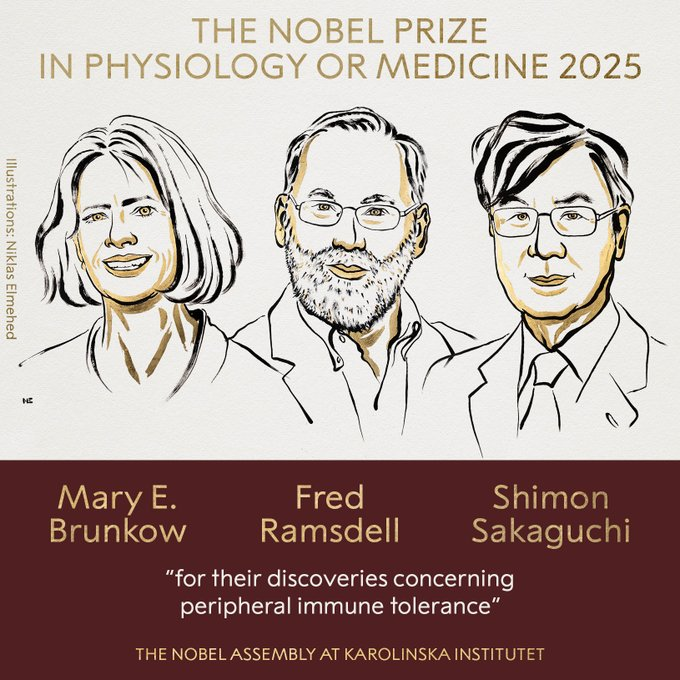 Ill. Niklas Elmehed © Nobel Prize Outreach
Today, let's explore this landmark discovery that transformed the field of immunology.
The Immune System's Dilemma
Our immune system protects us every day from countless viruses, bacteria, and other microorganisms. Without a functional immune system, we simply could not survive.
But here's the fundamental question: how does the immune system tell "friend" from "foe"?
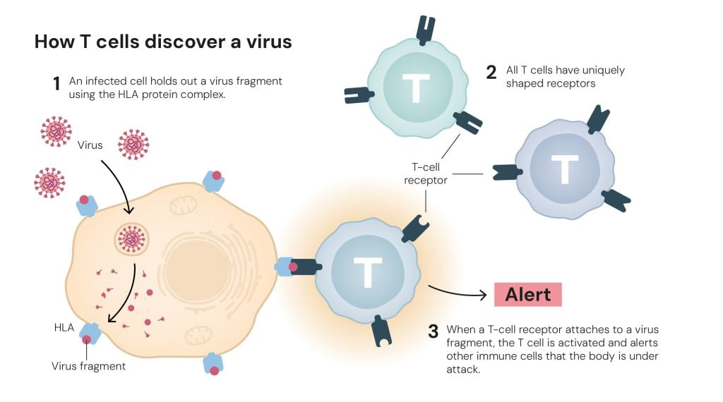 How T cells discover a virus. © The Nobel Committee for Physiology or Medicine. Ill. Mattias Karlén
To recognize as many pathogens as possible, our T cell receptors (TCRs) can generate more than 10^15 distinct receptor variants through V(D)J recombination. This remarkable diversity ensures we can recognize almost any foreign invader. However, it comes with an inherent cost — some self-reactive T cells capable of recognizing our own tissues are inevitably generated.
If these "rogue" T cells go unchecked, they attack our own organs, causing autoimmune disease. So how does the immune system maintain this delicate balance?
Central Tolerance: The Thymic Selection Mechanism
By the 1980s, scientists had already identified a mechanism called central tolerance. As T cells mature in the thymus, they undergo a rigorous "screening exam": T cells that strongly recognize self-proteins are eliminated through apoptosis ("negative selection"), and only those that pass the test are allowed to enter peripheral circulation.
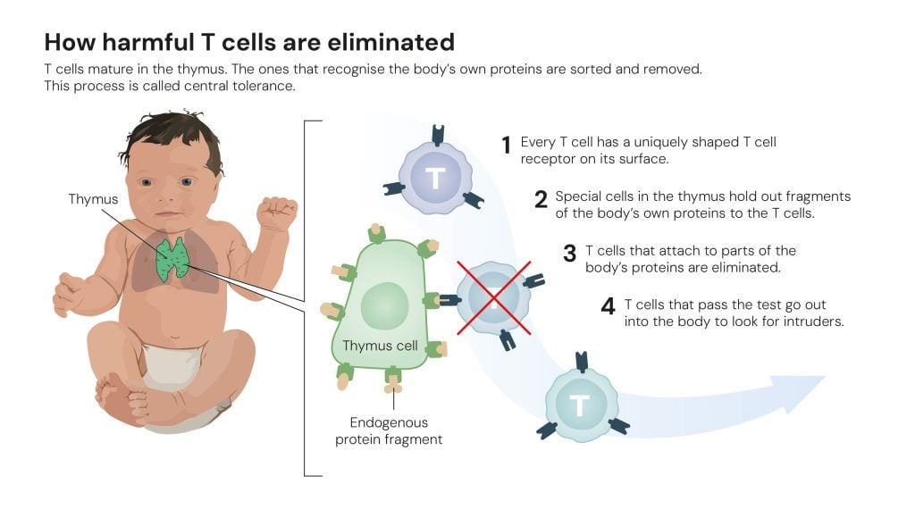 How harmful T cells are eliminated © The Nobel Committee for Physiology or Medicine. Ill. Mattias Karlén
This mechanism is critically important. In the early 2000s, scientists discovered that AIRE (autoimmune regulator), a transcription factor expressed in thymic medullary epithelial cells, drives the ectopic expression of tissue-specific antigens in the thymus — enabling the deletion of self-reactive T cells before they can escape. Mutations in the AIRE gene cause a rare autoimmune syndrome known as APS-1 (APECED).
But central tolerance is not perfect. Some self-reactive T cells inevitably slip through the thymic screen and enter peripheral tissues. So who keeps these escapees in check?
Sakaguchi's Perseverance: Discovering Regulatory T Cells
From Skepticism to Breakthrough
In the 1970s, the concept of "suppressor T cells" — a population capable of dampening immune responses — had been proposed. However, due to the imprecision of research methods at the time, many results proved unreproducible, and the entire field collapsed in the 1980s.
Yet Shimon Sakaguchi (坂口志文) did not give up.
In 1969, Nishizuka and Sakakura had shown that mice thymectomized at day 3 after birth spontaneously developed multiple autoimmune diseases, including oophoritis, gastritis, and thyroiditis.
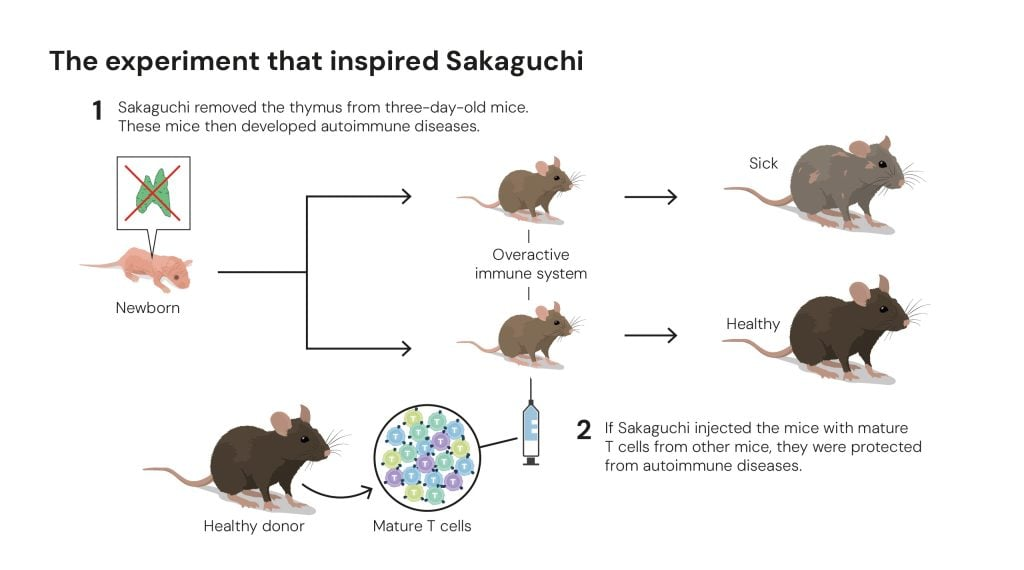 The experiment that inspired Sakaguchi © The Nobel Committee for Physiology or Medicine. Ill. Mattias Karlén
In 1982, while working in Nishizuka's laboratory, Sakaguchi found that transferring a specific subset of immune cells (CD5+CD45RB^low^) from normal mice into thymectomized mice prevented the development of autoimmune disease.
This pointed to a remarkable conclusion: the immune system must contain some kind of "safety guardian" capable of suppressing the activity of other T cells.
1995: Defining the Key Subpopulation
With advances in monoclonal antibody technology, Sakaguchi was now able to isolate T cell subpopulations with much greater precision. In 1995, he published a landmark paper in the Journal of Immunology demonstrating that:
- CD4+CD25+ T cells possess immunoregulatory function
- Depletion of these cells caused mice to spontaneously develop multiple autoimmune diseases
- Reconstitution with CD4+CD25+ T cells prevented disease
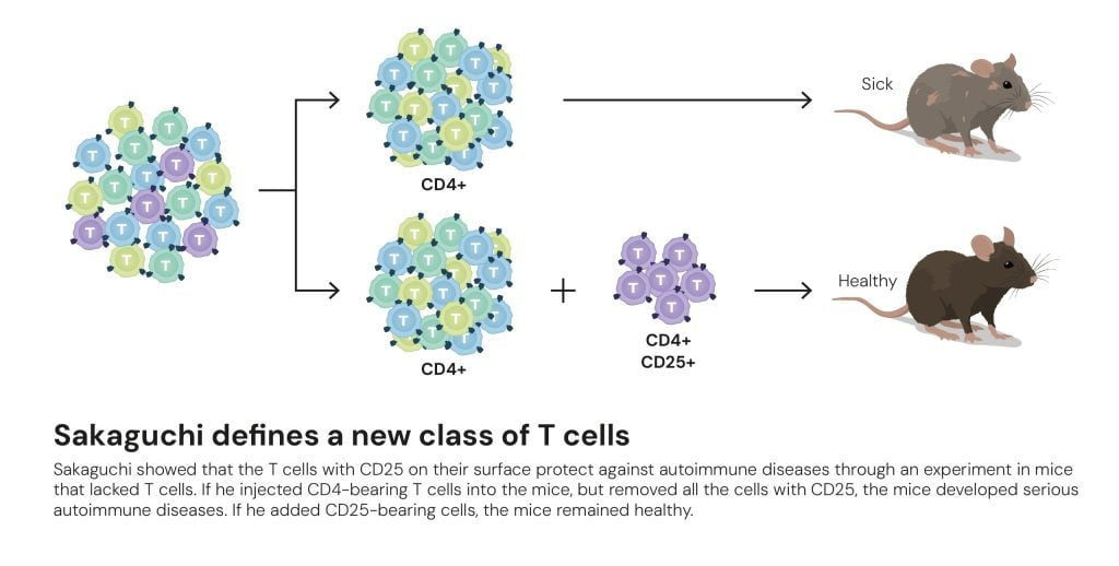 Sakaguchi defines a new class of T cells © The Nobel Committee for Physiology or Medicine. Ill. Mattias Karlén
These cells were named regulatory T cells (Tregs), establishing them as central players in peripheral immune tolerance.
Brunkow and Ramsdell: Cracking the FOXP3 Mystery
The Secret of the Scurfy Mouse
A parallel thread in this story begins with a mutant mouse strain called "scurfy" — named for its scaly, dandruff-like skin. This strain was serendipitously discovered in the 1940s at Oak Ridge National Laboratory in the United States, where scientists were studying the effects of radiation on mice.
Male scurfy mice die within weeks of birth, displaying severe systemic autoimmune manifestations: scaly skin, massively enlarged spleen and lymph nodes, and widespread multi-organ inflammation. Genetic analysis showed the lethal mutation mapped to the X chromosome.
Finding a Needle in a Haystack: Mapping the Foxp3 Gene
In the 1990s, Mary Brunkow and Fred Ramsdell were working at Celltech Chiroscience in the United States, focused on developing treatments for autoimmune diseases. They recognized that the scurfy mouse might hold an important clue.
They made a bold decision: find the mutated gene responsible for the scurfy phenotype.
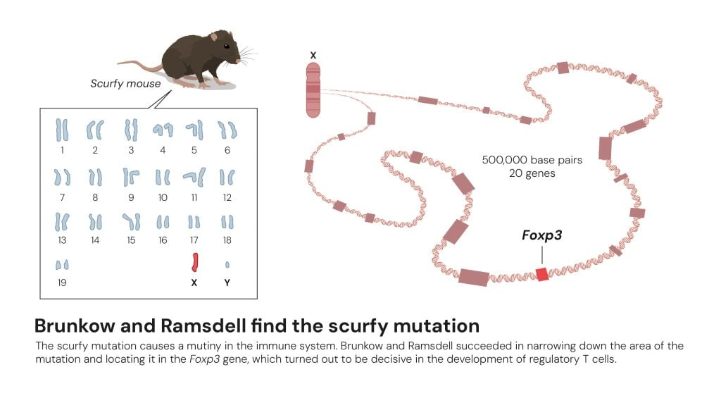 Brunkow and Ramsdell find the scurfy mutation © The Nobel Committee for Physiology or Medicine. Ill. Mattias Karlén
In those days, this was truly like finding a needle in a haystack. The mouse X chromosome spans approximately 170 million base pairs, and identifying a single causative mutation required enormous patience and a measure of good fortune. After years of painstaking effort, they:
- Narrowed the candidate region to approximately 500,000 base pairs
- Identified 20 candidate genes within that region
- Systematically compared gene sequences between healthy and scurfy mice
- Discovered a 2-base-pair insertion mutation in the very last gene they examined
This gene had never been described before. Because it shared homology with a gene family known as the "forkhead box" (FOX) family, Brunkow and Ramsdell named it Foxp3 (Forkhead box P3).
From Mouse to Human
Even more exciting was the realization that a rare human disease — IPEX syndrome (immune dysregulation, polyendocrinopathy, enteropathy, X-linked) — might be the human equivalent of the scurfy mouse condition.
Boys with IPEX syndrome develop fatal multi-organ autoimmunity shortly after birth, with involvement of multiple endocrine organs and the intestinal tract. Through collaborative work with pediatric clinicians worldwide, in 2001, Brunkow and Ramsdell published a paper in Nature Genetics demonstrating that:
- Mutations in human FOXP3 cause IPEX syndrome
- Patients with IPEX lack functional Treg cells
The Puzzle Complete: FOXP3 as the Master Regulator of Treg Development
In 2003, the final piece of the puzzle fell into place.
The Sakaguchi group and the Ramsdell group independently demonstrated almost simultaneously that FOXP3 is the master transcription factor governing Treg cell development and function:
- Foxp3 is specifically expressed in CD4+CD25+ Treg cells
- Ectopic expression of Foxp3 in conventional CD4+ T cells is sufficient to convert them into Treg cells
- Foxp3-deficient mice completely lack Treg cells and develop the same autoimmune phenotype as scurfy mice
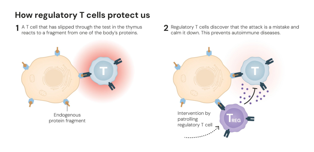 How regulatory T cells protect us © The Nobel Committee for Physiology or Medicine. Illustration: Mattias Karlén
At this point, a complete and coherent picture had emerged:
- The FOXP3 gene encodes a transcription factor
- FOXP3 controls the differentiation and function of Treg cells
- Treg cells suppress self-reactive T cells through multiple mechanisms
- Loss of functional FOXP3/Treg cells leads to severe, fatal autoimmune disease
How Treg Cells Work
How do Treg cells fulfill their role as "safety guardians"? Research has identified multiple suppressive mechanisms:
- Contact-dependent suppression: Direct inhibition of other T cells through surface molecules such as CTLA-4
- Secretion of inhibitory cytokines: Including IL-10, IL-35, and TGF-β
- IL-2 competition: Treg cells constitutively express high levels of the IL-2 receptor (CD25), enabling them to consume local IL-2 and effectively "starve" effector T cells
- Regulation of antigen-presenting cells: Modulating the maturation and function of dendritic cells
Clinical Applications
These fundamental discoveries have already catalyzed exciting clinical developments:
1. Autoimmune Disease Treatment
- Low-dose IL-2 therapy: Stimulates expansion of endogenous Treg cells
- Ex vivo Treg expansion and infusion: Being evaluated for type 1 diabetes and graft-versus-host disease (GvHD)
- CAR-Treg cells: Engineered Tregs designed to home to specific organs and prevent transplant rejection
2. Cancer Immunotherapy
- Anti-CCR8 antibodies: Selectively deplete tumor-infiltrating Tregs to enhance anti-tumor immunity
- Multiple clinical trials are currently underway
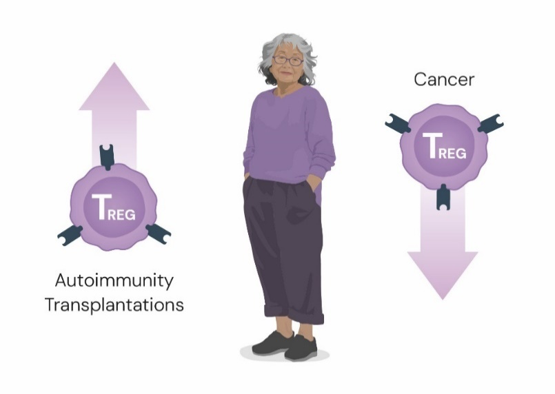 Approaches to harness or interfere with Treg cells in the treatment of disease. © The Nobel Committee for Physiology or Medicine.
3. Allergic and Inflammatory Diseases
- A 2024 randomized controlled trial demonstrated the efficacy of the IL-2 receptor agonist rezpegaldesleukin in moderate-to-severe atopic dermatitis
To date, more than 200 clinical trials are evaluating Treg cell-based therapies.
From Sakaguchi's unwavering persistence in the 1980s, to Brunkow and Ramsdell's painstaking positional cloning work in the 1990s, to the elucidation of FOXP3 function in the early 2000s — this story beautifully illustrates the power of fundamental scientific research: curiosity-driven inquiry, sustained effort, and the synergy of independent research teams converging on the same discovery.
The work of these three scientists not only answered a foundational question in immunology — how the immune system avoids attacking itself — but also opened transformative new paths for treating autoimmune diseases, cancer, and transplant rejection.
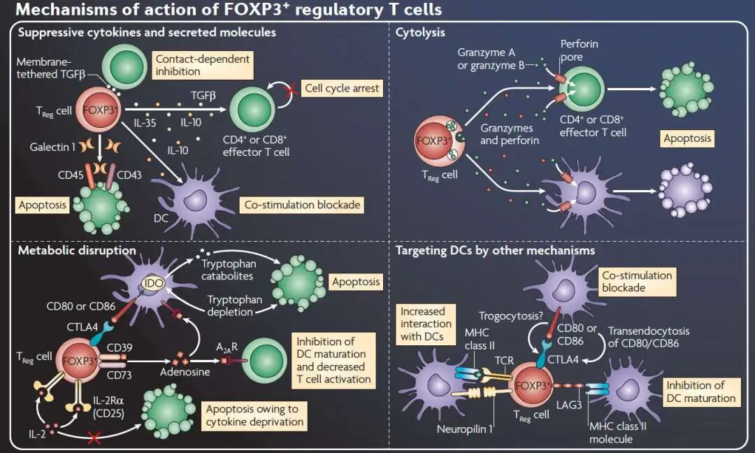 FOXP3 Treg function diagram. Shevach & Davidson, Nat Rev Immunology 2010
Appendix: Treg Cell Marker Gene Reference Table
Core Markers
| Marker Category | Protein | Human Gene | Mouse Gene | Expression | Function/Notes |
|---|---|---|---|---|---|
| Core triad | CD4 | CD4 | Cd4 | Positive | Helper T cell lineage marker |
| CD25 | IL2RA | Il2ra | High | IL-2 receptor α chain; defining Treg surface marker | |
| FOXP3 | FOXP3 | Foxp3 | Positive | Master transcription factor; most specific Treg marker | |
| Refined gating | CD127 | IL7R | Il7r | Low | IL-7 receptor; helps distinguish Tregs from activated conventional T cells |
Functional Markers
| Protein | Human Gene | Mouse Gene | Expression | Function |
|---|---|---|---|---|
| CTLA-4 (CD152) | CTLA4 | Ctla4 | Constitutive | Co-inhibitory molecule; critical for Treg suppressive function |
| GITR (CD357) | TNFRSF18 | Tnfrsf18 | High | TNF receptor superfamily member; activation marker |
| ICOS (CD278) | ICOS | Icos | Variable | Inducible T cell co-stimulator |
| TIGIT | TIGIT | Tigit | High | Inhibitory receptor |
| CD39 | ENTPD1 | Entpd1 | High in activated Tregs | Ectonucleotidase; converts ATP to AMP |
| CD73 | NT5E | Nt5e | High in activated Tregs | Ectonucleotidase; converts AMP to adenosine |
Subpopulation-Specific Markers
| Subpopulation | Protein | Human Gene | Mouse Gene | Expression | Notes |
|---|---|---|---|---|---|
| Thymus-derived Tregs (tTregs) | Helios | IKZF2 | Ikzf2 | Partial | Helps distinguish tTregs from peripherally induced pTregs |
| Neuropilin-1 | NRP1 | Nrp1 | High | Thymic origin marker | |
| Tumor-infiltrating Tregs | CCR8 | CCR8 | Ccr8 | Tumor Treg–specific high | Therapeutic target for tumor Treg depletion |
| LAG3 | LAG3 | Lag3 | High | Inhibitory receptor | |
| Effector Tregs | CD44 | CD44 | Cd44 | High | Activation/memory marker |
| KLRG1 | KLRG1 | Klrg1 | Positive | Terminal differentiation marker | |
| Central memory Tregs | CCR7 | CCR7 | Ccr7 | Positive | Lymph node homing receptor |
| CD62L | SELL | Sell | Positive | L-selectin; lymph node trafficking |
Activation State Markers
| State | Protein | Human Gene | Mouse Gene | FOXP3 | CD25 | CD45RA | Other Features |
|---|---|---|---|---|---|---|---|
| Resting Treg | CD45RA | PTPRC | Ptprc | Low | Low | Positive | Low suppressive activity |
| Activated Treg | — | — | — | High | High | Negative | CTLA4^high^ ICOS^high^ |
Nomenclature notes:
- Human gene symbols: all uppercase (e.g., FOXP3)
- Mouse gene symbols: first letter capitalized, remainder lowercase (e.g., Foxp3)
- Official HGNC gene names corresponding to CD antigens are indicated in the table
- Superscripts: ^high^ (high expression), ^low^ (low expression), ^+^ (positive), ^−^ (negative)
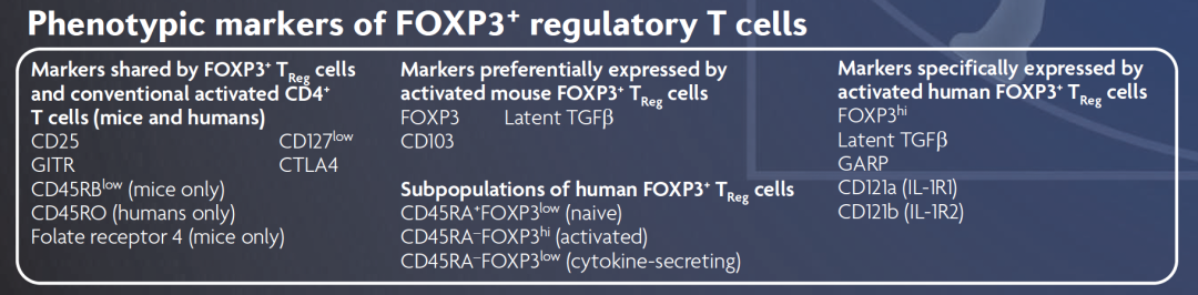 FOXP3+ Treg phenotype markers. Shevach & Davidson, Nat Rev Immunology 2010
References
- Popular information. NobelPrize.org. Nobel Prize Outreach 2025. Wed. 8 Oct 2025. https://www.nobelprize.org/prizes/medicine/2025/popular-information/
- Advanced information. NobelPrize.org. Nobel Prize Outreach 2025. Wed. 8 Oct 2025. https://www.nobelprize.org/prizes/medicine/2025/advanced-information/
- Shevach, Ethan, and Todd Davidson. "Regulatory T Cells." Nature Reviews Immunology, Macmillan Publishers, 2010. https://www.nature.com/documents/nri_posters_tregcells.pdf. Poster. Accessed 8 Oct. 2025.
Reply with nobel2025treg to receive the PDF of the key papers and the Nature Treg cell poster~
Originally posted on WeChat Official Account: bioinfo_studyroom