Paper Outlook — August 31 - September 6, 2026

TL;DR
- Spatial transcriptomics is maturing: SpaGCN uses graph neural networks to fuse gene expression, location, and histology, BayesSpace sharpens resolution with Bayesian neighborhood models, and G2T tries the reverse — predicting tissue layout from gene expression alone using generative flow matching.
- Single-cell and single-nucleus methods are being put to work on biological questions, not just method papers: from how obesity rewires estrogen signaling in endometrial tumors to whether the herbal compound berberine can suppress breast cancer by spurring mitochondrial cleanup.
- Manual marker-based cell typing is drawing more skepticism — the GmGM framework instead learns cell and gene relationships together, which may recover subtle cell states that conventional pipelines flatten.
- Multi-omics plus wet-lab validation is becoming standard for drug-mechanism work: public datasets, single-cell data, molecular docking, and bench experiments stitched together to nominate and confirm targets.
- Generative deep learning is creeping into spatial biology, pointing toward a shift from purely descriptive spatial analysis toward predicting or imputing tissue architecture where the spatial data is missing or too expensive to produce.
Single-nuclei RNA sequencing reveals obesity-associated rewiring of estrogen signaling in endometrioid adenocarcinoma.
This paper uses single-nucleus RNA sequencing to compare endometrial tumors from obese and normal-weight postmenopausal patients. The authors show that obesity reshapes estrogen-related gene networks not just in tumor cells but in the surrounding immune and stromal cells too, suggesting that obesity's link to this cancer involves more than hormone dosage. The work is a concrete example of how systemic body state can rewire a tumor microenvironment.
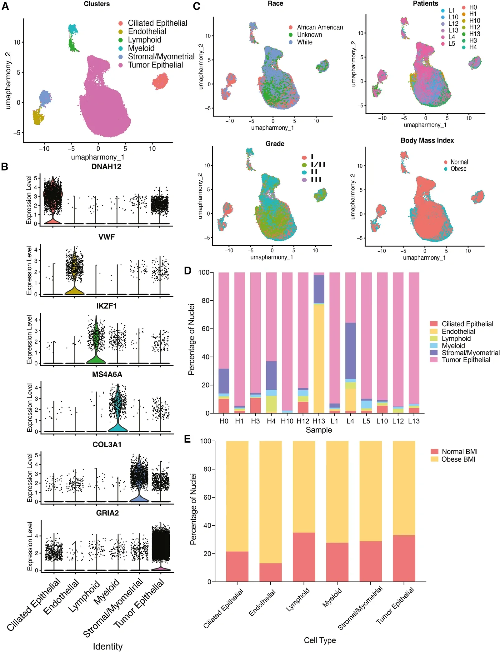
Figure 1 from Long JL et al., iScience (2026). Licensed CC BY-NC-ND. Source: 10.1016/j.isci.2026.117226.
iScience (2026)
Read the original → https://doi.org/10.1016/j.isci.2026.117226
Berberine Modulates Mitophagy-Related Gene Expression and Suppresses Breast Cancer Cells: A Multi-Omics and Experimental Study.
The authors combine public gene-expression datasets, single-cell data, molecular docking simulations, and lab experiments to argue that the natural compound berberine can suppress breast cancer cells by turning on mitophagy — the cell's process for clearing damaged mitochondria. The study points to specific genes through which berberine acts, giving plausible leads for follow-up drug work. It is a fairly clean example of multi-omics used end to end, from data mining to bench validation.
Cell biochemistry and biophysics (2026)
Read the original → https://doi.org/10.1007/s12013-026-02156-3
SpaGCN: Integrating gene expression, spatial location and histology to identify spatial domains and spatially variable genes by graph convolutional network
SpaGCN combines gene expression data, the physical location of each measurement spot on a tissue slide, and the slide's histology image to find spatial domains — regions where nearby cells share expression patterns and tissue appearance. It uses a graph neural network so each spot can borrow information from its neighbors. That makes it easier to pick out tissue structures and the genes whose expression varies across space.

Nature Methods (2021) · 1024 citations
Read the original → https://doi.org/10.1038/s41592-021-01255-8
Spatial transcriptomics at subspot resolution with BayesSpace
BayesSpace uses a Bayesian model to sharpen the resolution of spatial transcriptomics data, which is usually blurry because each measurement covers multiple cells. By treating neighboring spots as related, it effectively guesses what finer-scale expression patterns sit underneath the original resolution. The method also clusters spots into coherent tissue regions and has been tested on brain and melanoma samples, among others.
Nature Biotechnology (2021) · 892 citations
Read the original → https://doi.org/10.1038/s41587-021-00935-2
G2T: Tissue Reconstruction from Gene Expression via Embedding-Distance Flow Matching
G2T is a deep learning model that takes gene expression as its only input and predicts where cells likely sat within a tissue. It uses a Transformer architecture trained to predict pairwise distances between cells in a learned embedding space. The use case is the obvious gap: spatial data is missing or a cheaper experiment only produced single-cell profiles without coordinates.

bioRxiv (2026)
Read the original → https://doi.org/10.64898/2026.08.25.746917
Uncovering Cellular Resolution in scRNAseq via Unbiased Cell and Gene Network Analysis
This paper argues that standard single-cell RNA-seq analysis leans too heavily on manual marker genes, and that this can miss subtle cell states. The authors apply a statistical framework that learns which genes depend on each other and which cells are similar at the same time, producing more stable and unbiased clusters. On a blood cell dataset, the approach recovered cell types matching expert annotation without any manual gene selection.
arXiv (2026)
Read the original → https://arxiv.org/abs/2608.22982v1
Quick links
| Paper | Venue / Year | Link |
|---|---|---|
| Single-nuclei RNA sequencing reveals obesity-associated rewiring of estrogen signaling in endometrioid adenocarcinoma. | iScience (2026) | Read → |
| Berberine Modulates Mitophagy-Related Gene Expression and Suppresses Breast Cancer Cells: A Multi-Omics and Experimental Study. | Cell biochemistry and biophysics (2026) | Read → |
| SpaGCN: Integrating gene expression, spatial location and histology to identify spatial domains and spatially variable genes by graph convolutional network | Nature Methods (2021) | Read → |
| Spatial transcriptomics at subspot resolution with BayesSpace | Nature Biotechnology (2021) | Read → |
| G2T: Tissue Reconstruction from Gene Expression via Embedding-Distance Flow Matching | bioRxiv (2026) | Read → |
| Uncovering Cellular Resolution in scRNAseq via Unbiased Cell and Gene Network Analysis | arXiv (2026) | Read → |