Journal Club: Multi-Omics Integration Across the Cancer Continuum – From Early Detection to Tumor Evolution
Introduction
Cancer is no longer viewed as a monolithic disease but as a complex ecosystem shaped by molecular heterogeneity, dynamic evolution, and constant crosstalk with its microenvironment. That complexity has made single-omics approaches — relying only on genomics, transcriptomics, or imaging — insufficient for precise diagnosis, prognosis, and treatment selection. The field is moving toward integrated multi-omics, a paradigm that stacks diverse molecular and clinical data types to build a more complete picture of a tumor.
This month we look at three recent studies from Nature Communications that span different stages of the cancer journey. Together they show how multi-omics is being put to work on real clinical problems: the diagnostic uncertainty of early lung nodules, the cellular plasticity driving pediatric liver tumors, and the molecular subtyping of advanced lung cancers. Each paper layers a different combination of omics — clinic-radiomic-fragmentomic, single-cell transcriptomic-epigenomic-spatial, and proteogenomic — matched to its specific biological and clinical question.
The bigger picture is a shift from descriptive biology to clinically integrated models. Multi-omics signatures are starting to guide non-invasive diagnoses, reveal how tumors resist therapy, and redefine cancer classifications beyond what a microscope can see.
Paper 1: A Multi-Omics Model for Diagnosing Indeterminate Lung Nodules
Key Findings
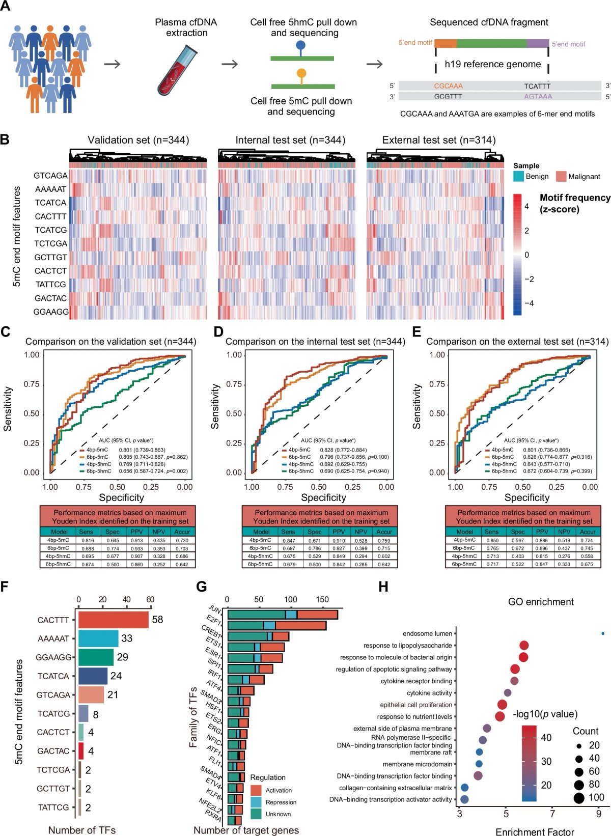
- Diagnostic accuracy: The integrated clinic-RadmC model — combining clinical data, CT radiomics, and cfDNA (circulating cell-free DNA) fragmentomics in 5mC-enriched regions (5mC = 5-methylcytosine, an epigenetic DNA mark) — hit an AUC (area under the receiver-operating-characteristic curve, a standard accuracy metric from 0 to 1) of 0.923 on an external test set of 2032 participants. That beat any single-omics model or partial combination tested (p < 0.05).
- Clinical impact: The model could, in theory, reduce unnecessary invasive procedures for benign nodules by 10.9–35% and avoid delayed treatment for malignant nodules by 3.1–38.8%.
- Robustness: The gain over standard clinic-radiological assessment held after adjustment for those variables, suggesting clinic-RadmC adds independent information on top of what clinicians already measure.
- Why methylation-guided fragmentomics: The team focused cfDNA fragmentation patterns on genomic regions enriched for 5mC rather than the whole genome. That targeted view was more informative, hinting at a link between tissue-specific methylation and the fragmentation signatures cancers shed into blood.
Methodology & Study Design
- Technical approach: A multi-institutional diagnostic study. Three data layers were integrated: (1) clinical features such as age and nodule characteristics, (2) radiomic features from chest CT capturing nodule texture, shape, and intensity, and (3) fragmentomic features from low-coverage whole-genome sequencing of plasma cfDNA, looking at fragment size and end motifs in 5mC-enriched regions.
- Cohort: 2,032 participants with indeterminate pulmonary nodules (IPNs), split into training, validation, and external test sets.
- What's new: Earlier models had paired clinical + radiomic or clinical + cfDNA data. This one stacks all three into a single, externally validated framework, and it does so with a methylation-targeted fragmentomics layer that hadn't been used at this scale.
Significance
Indeterminate pulmonary nodules are common, and working them up is expensive, invasive, and anxiety-inducing. A blood-plus-scan test that sorts benign from malignant with high accuracy would be a meaningful win. The broader lesson is methodological: the synergy between imaging, liquid biopsy, and clinical data lands further than any layer alone, and it sets a template for how integrated diagnostic models should be built and validated.
Paper 2: Single-Cell Multi-Omics Maps Plasticity and Clonal Evolution in Hepatoblastoma
Key Findings
- A continuum of cell states: Single-cell multi-omics showed hepatoblastoma (HB) cells sitting along a continuum of differentiation states, anchored by hepatocytic (scH), liver progenitor (scLP), and mesenchymal (scM) poles. A new intermediate scH/LP population was found at the spatial border between scH and scLP regions on the spatial transcriptomics map.
- Clonal plasticity: By assigning somatic mutations to single cells, the authors showed that each genetic subclone occupies its own range of cell states. So genetic evolution (clonality) and non-genetic phenotypic diversity (plasticity) are entangled, not separate.
- Therapy response: scLP-dominant subclones, which overexpressed stem cell and DNA repair genes, proliferated faster after neoadjuvant chemotherapy (treatment given before surgery to shrink the tumor). That points to a progenitor-like, plastic state as a likely reservoir for resistance and relapse.
- Regulatory wiring: Single-cell ATAC-seq (Assay for Transposase-Accessible Chromatin sequencing — a readout of which parts of the genome are open and likely active in each cell) mapped the gene regulatory networks and transcription factor programs that drive transitions between these states.
Methodology & Study Design
- Technical approach: A single-cell multiomic workflow on primary HB samples that co-assays the transcriptome (scRNA-seq) and chromatin accessibility (scATAC-seq) from the same cells, combined with bulk whole-genome sequencing for somatic mutations and spatial transcriptomics to place cell states within tumor architecture.
- Core innovation: Capturing genotype (mutations per clone), phenotype (transcriptional state), and epigenotype (chromatin accessibility) at single-cell resolution gives a much sharper view of how genetic subclones move through a space of possible cellular identities.
Significance
This paper goes beyond cataloging heterogeneity — it explains where it comes from and what it does. The central argument is that clonal evolution and epigenetic plasticity act together to drive HB biology and clinical behavior. By flagging the scLP state as chemoresistant, the work reframes the target: rather than chasing a static "cancer cell," it points toward the dynamic processes that let cells switch states and adapt, which has clear implications for designing therapies that lock cells in a sensitive state or clear out the plastic progenitor pool.
Paper 3: Proteogenomic Subtyping Reveals NSCLC Biology Beyond Histology
Key Findings
- Five molecular subtypes: Integrative analysis of 229 Korean NSCLC (non-small cell lung cancer) patients, plus 462 from public cohorts, defined five molecular subtypes that cut across the traditional histological split between LUAD (lung adenocarcinoma) and LSCC (lung squamous cell carcinoma). One subtype, marked by PI3K-Akt pathway upregulation, had high metastasis rates and poor survival regardless of histology.
- Chromosome instability link: Proliferative subtypes in both LUAD and LSCC tracked strongly with whole genome doubling (WGD) — a major driver of chromosomal instability (CIN) and tumor aggressiveness.
- Diverse immune microenvironments: Immune cell composition and neoantigen load (the repertoire of novel peptides a tumor presents to the immune system) varied widely across subtypes. Distinct immunological subtypes emerged, including a "hot" tumor-enriched state that responded better to adjuvant therapy (additional treatment given after the main therapy to lower the chance of recurrence).
- Toward clinical translation: The framework connects molecular features to actionable clinical traits — CIN for aggressiveness and possible therapeutic vulnerabilities, and tumor microenvironment composition for predicting immunotherapy response.
Methodology & Study Design
- Technical approach: A proteogenomic study integrating whole-exome sequencing, RNA sequencing, and mass spectrometry-based proteomics on tumor tissue. That stack links genomic alterations to their functional protein-level consequences and to immune context.
- Cohort: A sizable, uniformly processed Korean discovery cohort, validated against large public multi-omics resources (TCGA, CPTAC).
Significance
Lung cancer classification has leaned on histology for over a century. This work argues that molecular subtypes defined by proteogenomic integration capture something more useful for prognosis and treatment selection — CIN, immune context — than microscopic appearance alone. It offers a concrete blueprint for a next-generation, biology-driven taxonomy of NSCLC that could shape clinical trial design and personalized therapy.
Synthesis & Discussion
Taken together, these three papers tell a coherent story about multi-omics across the cancer continuum. Paper 1 sits at the diagnostic frontier, stacking clinical, imaging, and plasma DNA data to answer a binary question: benign or malignant? Its strength is near-term clinical translatability. Paper 3 sits at the subtyping and prognostic stage, using deep proteogenomic profiling of resected tumors to refine classification and predict behavior. Paper 2 dives into the mechanistic core of tumor biology, using the highest-resolution tools available — single-cell multi-omics — to explain how heterogeneity and therapy resistance arise from the interplay of clones and plasticity.
The three are complementary rather than overlapping. The clonal plasticity seen in HB (Paper 2) is plausibly the engine that generates the heterogeneous subtypes captured in NSCLC (Paper 3), whose traces can be picked up non-invasively by the fragmentomic layer in Paper 1. A pattern that emerges across all three is the move from static classification to dynamic mapping: from labeling a tumor as a type (LUAD) to understanding it as a system of co-existing cell states (Paper 2), which gives rise to a dominant molecular phenotype (Paper 3), whose signals can be detected in blood (Paper 1).
No direct contradictions, but a real tension runs through the set: scale versus resolution. Papers 1 and 3 use bulk or liquid-biopsy analyses on large cohorts — necessary for biomarker development and clinical validation. Paper 2 gives unmatched resolution but on a smaller scale, and it's not yet clear how to lift single-cell mechanistic insights into clinical-grade assays. The likely path forward is to use single-cell studies like Paper 2 to nominate the features that bulk or liquid assays like Papers 1 and 3 should aim to capture.
Open questions worth keeping in mind:
- Technical: How do we standardize data integration pipelines across platforms and institutions so results reproduce?
- Biological: Does the plasticity continuum described in pediatric HB (Paper 2) hold up in adult carcinomas like NSCLC?
- Clinical: Will the diagnostic AUCs reported in Paper 1 translate into fewer invasive procedures and lower mortality in prospective, randomized trials?
For Your Lab Meeting
Here are 5 questions to spark discussion:
- Methodology: Paper 1 analyzed cfDNA fragmentomics in 5mC-enriched regions. What are the trade-offs of that targeted approach versus a genome-wide fragmentomics analysis?
- Generalizability: Paper 3's molecular subtypes were derived from a Korean cohort. How might population-specific genetic backgrounds or environmental exposures shift the prevalence or behavior of these subtypes? What would global validation actually require?
- Future directions: Given the clone-and-plasticity interplay in Paper 2, what would a therapy designed to constrain tumor cell plasticity (rather than just kill proliferating cells) actually look like?
- Connecting the dots: If you were designing a study that follows a lung nodule from detection (Paper 1's domain) through to resection and molecular profiling (Paper 3's domain), what longitudinal multi-omics data would you collect to bridge the two and capture tumor evolution?
- Broader trends: All three papers rely on heavy computational integration. What are the biggest unsolved problems in bioinformatics and machine learning for multi-omics data fusion, and which emerging tools or algorithms look most promising?
Key Terms
- Fragmentomics: The study of patterns in cell-free DNA fragments — length, genomic end positions, coverage imbalances. These patterns reflect the chromatin structure of the cells of origin and can act as non-invasive cancer biomarkers.
- Proteogenomics: An integrated analysis combining genomic, transcriptomic, and proteomic data. It looks for protein-level evidence of genomic alterations (for example, novel peptides produced by mutations) and for cases where mRNA expression does or doesn't translate into protein.
- Cellular plasticity: The ability of cancer cells to switch between phenotypic states (stem-like, differentiated, mesenchymal, and so on) in response to environmental cues or treatment. It is a non-genetic, often epigenetically regulated, driver of tumor heterogeneity and therapy resistance.
- Radiomics: High-throughput extraction of quantitative features — texture, shape, intensity — from medical images. Many of these features are invisible to the eye but can feed predictive models for diagnosis, prognosis, or treatment response.
- Whole Genome Doubling (WGD): An event in which a cell ends up with two full copies of its genome. WGD is a major source of chromosomal instability, supplies raw material for further evolution, and is tied to poor prognosis in many cancers.
References
Mengmeng Zhao, Gang Xue, Bingxiang He, J. Deng, Tingting Wang, Y. Zhong, et al. (2025). Integrated multiomics signatures to optimize the accurate diagnosis of lung cancer. Nature Communications. https://doi.org/10.1038/s41467-024-55594-z
Amelie Roehrig, T. Hirsch, Aurore Pire, G. Morcrette, Barkha Gupta, C. Marcaillou, et al. (2024). Single-cell multiomics reveals the interplay of clonal evolution and cellular plasticity in hepatoblastoma. Nature Communications. https://doi.org/10.1038/s41467-024-47280-x
K. J. Song, Seunghyuk Choi, Kwoneel Kim, H. S. Hwang, E. Chang, Ji Soo Park, et al. (2024). Proteogenomic analysis reveals non-small cell lung cancer subtypes predicting chromosome instability, and tumor microenvironment. Nature Communications. https://doi.org/10.1038/s41467-024-54434-4